Table of Contents
Advertisement
Quick Links
Advertisement
Table of Contents

Related Manuals for iCR iCR 3600


Summary of Contents for iCR iCR 3600
- Page 1 iCR3600+ USER MANUAL...
- Page 2 All other trademarks are the property of their respective owners, and are hereby acknowledged. Terms Any one of the following iCR products will be referred to as the “CR unit” throughout this document: iCR 1000 , iCR 1000 Dual...
-
Page 3: Contact Information
31a Ridlerstrasse, 80339 Munich, Germany Phone: +49.89.2555.757.150 Email: info@icrcompany.com Web: http://www.icrcompany.com iCR 3600+ Information Please Enter the details of the iCR 3600+ system here: Serial Number: Date Purchased: Interface Type: USB 2.0 c 2007-2009 ii of 69 Document # 3600+-01A Rev C Confidential and Proprietary... -
Page 4: Safety Information
3600 + – User Manual Safety Information Be sure to read and understand the installation and operating instructions before applying power to the CR unit. C A U T I O N INPUT: 100-240VAC 3A 50-60Hz CAUTION: FOR CONTINUED... - Page 5 3600 + – User Manual replaced by properly trained service personnel. Contact iCRco if there are any issues with the covers being damaged or replacement covers are needed. Electrical Hazards WARNING This equipment is operated with hazardous voltages which can shock, burn, or cause death.
- Page 6 Guidance and manufactures’ declaration – electromagnetic emissions The iCR 3600+ is intended for use in the electromagnetic environment specified below. The customer or the user of the iCR 3600+ should assure that it is used in such an environment. Emissions test Compliance Electromagnetic environment –...
- Page 7 ) for 0.5 ) for 0.5 hospital environment. If the user tions and cycle. cycle. of the iCR 3600+ requires voltage continued operation during variations 40% U (60% 40% U (60% power mains interruptions, it is...
- Page 8 RF IEC to 80 MHz communications equipment 61000-4-6 should be used no closer to any 3 V/m 80 MHz 3 V/m part of the iCR 3600+, including Radiated to 2.5 GHz cables, than the recommended RF IEC separation distance calculated 61000-4-3 from the equation applicable to the frequency of the transmitter.
-
Page 9: Icrco Warranty
To assess the electromagnetic environment due to fixed RF transmitters, an electromagnetic site survey should be considered. If the measured field strength in the location where the iCR 3600+ is used exceeds the applicable RF compliance levels above, the iCR 3600+ should be observed to verify normal operation. - Page 10 3600 + – User Manual the media, not including hard drives, on which the software is furnished will be free from defects in materials and workmanship under normal use for a period of one (1) year from the date of original shipment from iCRco. iCRco’s sole obligation under this warranty is...
- Page 11 3600 + – User Manual customer name and contact information; product location and operating conditions; a copy of the purchase documents, and sufficient information and authorization, including a liabil- ity release as to any loss of data (that should always be backed up), software or network injury, or downtime, allowing iCRco technicians remote access to the product.
-
Page 12: Revision History
3600 + – User Manual ARE LIMITED IN DURATION TO THE INITIAL WARRANTY PERIOD AND NO WAR- RANTIES, EXPRESS OR IMPLIED, WILL APPLY AFTER THIS PERIOD. ALL INFORMA- TION, SPECIFICATIONS, PRICES, AND SERVICES ARE SUBJECT TO CHANGE AT ANY TIME WITHOUT NOTICE. -
Page 13: Table Of Contents
Periodic Cleaning ........5.6.1 Cleaning the Outside of the iCR 3600+ ....c 2007-2009... - Page 14 3600 + – User Manual 5.6.2 Cassette Cleaning ......6 QPC XSCAN32 Software Functions Creating/Editing Patients .
-
Page 15: Introduction
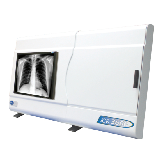
3600 + – User Manual 1. Introduction 1.1 Overview The CR unit is an ultra high resolution Computed Radiography device. It is designed to scan cassettes containing phosphor screens (CR plates) using patented technology. CR plates are transported past a scanning head without bending or using rollers. This flat scan path results in ultra high resolution images with high fidelity across the entire image. -
Page 16: Pre-Installation
2.2 Environmental The iCR 3600+ should not be placed in a room with a film processor present. This will void the warranty. The humidity and temperature limits are 20 to 80% non-condensing, and 50 to 95 F (10 to 35 C) operating, respectively. -
Page 17: Power Switch Location
While the unit is scanning the yellow light will blink. 2.6 Physical Requirements The iCR 3600+requires a stable operating environment. It is important the system is placed on a table, stand or wall mount that provides adequate support. 2.7 System Specifications... - Page 18 3600 + – User Manual Scan Rate Vibration/Acceleration Scan Rate 60 lines/second 3-4G Max (in shipping) Grey Scale Resolution Altitude 16 bits (65,535 shades of gray) Interface 0 to 9,500 ft. - operating USB 2.0 Weight Dimensions iCR 3600+...
-
Page 19: Hardware Installation
4. Store the box and any foam inserts somewhere safe & dry, so that if the iCR 3600+ needs to be shipped again, there are packing materials available. -
Page 20: International Power Cable
– User Manual 3.2.1 International Power Cable The iCR 3600+utilizes an international IEC grade connector for the power cable. Systems are shipped with a standard NEMA 5-15 hospital grade cable. The cable needs to be changed depending on the male end, which varies from country to country. - Page 21 3600 + – User Manual Figure 3.2: 4. Carefully slide the computer into the computer mounting bracket (Figure 3.3). Note For easy access to the CD-ROM drive, make sure the drive opens towards the outside of the unit. Figure 3.3: 5.
-
Page 22: Wall Mounting Plates
3600 + – User Manual Figure 3.4: 3.5 Wall Mounting Plates Notice Wall mounting plates must be installed by a licensed contractor. c 2007-2009 8 of 69 Document # 3600+-01A Rev C Confidential and Proprietary March 22, 2010 Property of iCRco, Inc. - Page 23 3600 + – User Manual 1. Attach the large wall mounting plate against wall at desired height. Allow the contractor to determine the proper screws to fasten the large wall mounting plate to the wall. 2. Attach the small wall mounting plate to the CR unit with three (3) 6-32 x ”...
-
Page 24: Software Installation
3600 + – User Manual 4. Software Installation 4.1 USB Driver Installation Installation of USB drivers are automatic with the installation of the iCRco software (i.e., QPC XSCAN32). 4.2 Installing QPC XSCAN32 QPC XSCAN32 software comes bundled with the CR unit. This software package will allow the user to interface with the CR unit. - Page 25 3600 + – User Manual 5. Make sure both QPC XSCAN32 Software and Software Key Driver boxes are checked, then click Next. 6. Select Scanning Station or Reading Station depend on the use, then click Next to con- tinue.
- Page 26 3600 + – User Manual 9. The Destination Folder should be set to C: Xscan32 then click Next to continue. 10. The Program Folder name should be set to QPC XSCAN32, then click Next. 11. QPC XSCAN32 will begin to install. Be patient while XSCAN32 installs.
-
Page 27: System Operation
2. Make sure the power cord is properly installed in the UPS and the UPS is plugged into a standard wall outlet. 3. Make sure the USB 2.0 cord is properly installed in the iCR 3600+ and computer. 4. Power on the computer. -
Page 28: Cassette Contamination
3600 + – User Manual 5.2.4 Cassette Contamination Should the situation arise where a cassette may come in contact with bodily fluids or other contaminated materials, place the cassette in a clean plastic bag before exposing the X-ray. This will ensure that the cassette stays clean and usable. -
Page 29: Acquiring An Image
3600 + – User Manual 4. Gently push the top of the cassette into place. 5. If loaded correctly, the user will see the white guide lines around the edge of the cassette. 6. Close the bay door. 5.4 Acquiring an Image WARNING This equipment employs a laser. - Page 30 6. Click on Scan to bring up the Quality Processing Center Scan dialog. (if it is a patient with no images the scan dialog will automatically appear). 7. The iCR Quality Processing Center dialog will appear. c 2007-2009 16 of 69 Document # 3600+-01A Rev C Confidential and Proprietary...
- Page 31 3600 + – User Manual 8. Select the anatomy by mousing over the skeleton on the left hand side of the Quality Processing Center dialog. 9. Select the cassette size by clicking the correct button (i.e. 14x17 in, 10x12 in). This will start the scan.
- Page 32 3600 + – User Manual 13. Remove any collimation by using the Auto Crop(located in the main toolbar), Mask, or User Mask (located in the Image Processing dialog) functions. 14. Process using ICE or ICE2 (located in the Image Processing dialog).
- Page 33 3600 + – User Manual 17. Return to the Patient Information window. Select the patient(s) to send to a PACS or viewing station. If you wish to send more than one patient, hold the Ctrl key down and left mouse click to select multiple patients.
-
Page 34: Schedule Of Maintenance
CAUTION Do not clean the galvo mirror. Dust and fibers in the laser beam path may affect the radiographic image. The outside covers of the iCR 3600+ should be cleaned with a slightly dampened cloth or a dry cloth moistened with Ball SUNUP glass cleaner or Sprayway c glass cleaner. -
Page 35: Cassette Cleaning
3600 + – User Manual 5.6.2 Cassette Cleaning CAUTION At no time should abrasive cleaners or chemicals be used to clean the cassette or plate. Cleaning the Outside of the Cassette 1. Moisten a clean, lint-free cloth with a mild soap or detergent using soft water. - Page 36 3600 + – User Manual Note If iCRco Plate Cleaner is not available, please contact Technical Support at 1-310-921-9559 to obtain more. 4. Gently wipe down the imaging plate with the clean, lint-free cloth. 5. Allow the plate to air dry before sliding the carbon fiber door back into place.
-
Page 37: Qpc Xscan32 Software Functions
3600 + – User Manual 6. QPC XSCAN32 Software Functions 6.1 Creating/Editing Patients 1. Open XSCAN, then click the New button in the Patient Information dialog. Alterna- tively, if the patient is already in the patient list, select the patient and click the Edit button on the Patient information dialog. - Page 38 3600 + – User Manual 1. Shows the currently selected anatomy 2. Shows the current patient name & birth date 3. Close: This button will close the scan interface (it will not close XSCAN) 4. Reset: This button will return the CR unit to the “home” position 5.
-
Page 39: Mammography Configuration
3600 + – User Manual 12. Scan preview: once the user has begun the scan, a preview image will appear as the plate scans. 6.3 Mammography Configuration Note The Mammography module is an add-on module. It may not be available in every country. - Page 40 3600 + – User Manual Notice POTENTIAL LOSS OF DATA! Be sure to take note of the current settings before entering the new values. The values will be overwritten and there is no way to recover them. The user will have to manually switch between settings when switching between general radiography and mammography.
-
Page 41: Default Mouse Functions For Image Manipulation
3600 + – User Manual 7. Click Ok again in the CR Settings window to save the settings. 6.4 Default Mouse Functions for Image Manipulation Windows / Level Images The middle mouse button is used to window / level images. Hold down the middle mouse... -
Page 42: Image Processing Dialog Box
3600 + – User Manual 6.5 Image Processing Dialog Box Window / Level Holding the middle mouse button (simultaneously holding the CTRL key and right mouse clicking will also window/level) down and moving the mouse vertically will adjust the level and horizontally will adjust the window. - Page 43 3600 + – User Manual Left and Right CR Markers 1. Open an image for viewing. 2. Select the Left/Right Markers button to apply a Left or Right marker to the image. 3. Select either the Left or Right marker. When the user clicks on the Left/Right Marker button the Left marker icon will appear.
- Page 44 3600 + – User Manual Before After CR Masking 1. Open an image for viewing. 2. Click the Mask button. 3. To remove the collimation mask, select the Mask button again. CR Masking Example Before Masking After Masking CR User Masking 1.
- Page 45 3600 + – User Manual 4. Hold the left mouse button down on the start region and move the mouse to the end of the mask region. 5. Repeat step 4 for each exposure. After the user has completed selecting the number of exposures, the masks will appear on the image.
- Page 46 3600 + – User Manual CR Processing 1. Open an image for viewing. 2. Select an anatomy from the CR Preset box. 3. Click the Process button to begin the processing. 4. The user may press the Reset button to restore the original image data set.
-
Page 47: Annotations
3600 + – User Manual Clicking the drop down menu button located below the “sharp” option gives the user the ability to refine the image further. After the user clicks on the drop down menu, an interface appears showing two sliders. The first slider enhances edges and contrast as the slider is moved to the right. - Page 48 3600 + – User Manual 3. Move the mouse to where the second endpoint will be. 4. Left mouse click on the are of the image to set the second endpoint. Extended Line Annotation Creates a straight line that passes through the image.
- Page 49 3600 + – User Manual 2. Hold down the left mouse button on the part of the image that is desired to start drawing then drag the mouse. 3. Continue to hold the left mouse button and draw as necessary .
- Page 50 3600 + – User Manual Measurement Annotation Used to measure the distance of a straight line on the image. This can be in inches, cm, mm and pixels. 1. Select the Measurement button from the annotations toolbar. 2. Left mouse click on the image to set the first endpoint of the measurement.
- Page 51 3600 + – User Manual Cobb Angle Annotation Used to draw a cobb angle on the image. 1. Select the Cobb Angle button on the annotations toolbar. 2. Left mouse click on the area of the image to set the first endpoint of the first line.
- Page 52 3600 + – User Manual 4. Left mouse click on the area of the image to set the third point. 5. Left mouse click on the area of the image to set the fourth point. The center line of these four points will be drawn on the image.
- Page 53 3600 + – User Manual Templates Used to load and draw custom templates. Select the Templates button on the annotations toolbar. Measurements Units Used to toggle between the current measurement units. Delete All Annotations Used to permanently remove all annotations from the currently selected images.
-
Page 54: Patient Cd/Dvd Creator
3600 + – User Manual 2. Move the mouse over the annotation onto any part of it except the handles (i.e. small boxes). The icon will change to the moving annotations icon. 3. Left mouse click on the annotations and drag it anywhere on the image. - Page 55 3600 + – User Manual Note If this is the first time using the patient CD creator feature a prompt will appear indicating the user to browse for the location of the DVD/CD Creator software. Alternatively, if Nero software is already installed on the PC, select the option Use Nero scripting to create CD/DVD.
- Page 56 3600 + – User Manual Using other CD/DVD recording software 1. When the software has finished initializing and copying the patient CD, a ‘Ready to create CD’ prompt will appear, select Yes. 2. The DVD/CD Creator software will open. Select source folder C: XscanCDFolder and drag the contents of the Xscan CD Folder folder and two files into the data project...
-
Page 57: Associating A Barcode With A Study
3600 + – User Manual 6.8 Associating a Barcode with a Study 1. In the patient information window, enter patient and study information. 2. Select patient and study. 3. Select Add Barcode button. 4. Using a barcode reader, scan the barcode of an unexposed cassette. -
Page 58: Finding Barcodes And Scanning Images
3600 + – User Manual 6.9 Finding Barcodes and Scanning Images 1. In the patient information window, select the Find Barcode Edit box. 2. Using a barcode reader, scan the barcode of the exposed cassette. 3. Select the anatomy information for the study. -
Page 59: Archiving Qpc Xscan32 Database
3600 + – User Manual The image below indicates that the image will appear under the patient Joe Smith and the barcode for the cassette that will be scanned is 01003. There is an option in the scan interface settings that requires the user to scan the barcode before they are able to scan cassettes into the interface. -
Page 60: Restoring Archived Database
3600 + – User Manual 5. When archiving is completed a message will appear informing the user of the archive location. Note the current date and time is used for the folder name of the archived database. 6.11 Restoring Archived Database WARNING It is strongly recommended to backing up data to non-spinning media CD/DVD or an archive (PACS) with RAID capability to ensure no loss of data. - Page 61 3600 + – User Manual 2. To search a particular archive, select the archive location by selecting the Browse button. 3. Under Date/Time, select from the list of valid archives 4. The user can either: (a) Select Find to open the entire database.
-
Page 62: Modality Work List Query
3600 + – User Manual 6.13 Modality Work List Query 6.13.1 Setting-up Modality Work List Query 1. Open QPC XSCAN32, then go to Options Preferences Image Transfer tab. 2. Check the box for Use DICOM Modality Work List Query. -
Page 63: Using Modality Work List Query
3600 + – User Manual Note The Text File Listener should only be used if the EMR/RIS/HIS does not support DICOM Modality Work List Query or HL7 Messaging. 1. In the XSCAN32 Modality Work List Query window, click the Options button. -
Page 64: Report Writer
3600 + – User Manual query by AET query by Date (i.e. today’s date only) (d) Click here to select the target to query. Note The user will only need to do this once. 3. The queried patients will appear in the patient list. Click on Retrieve Selected Patient(s) button to retrieve the patients into XSCAN32 database. -
Page 65: Long Bone & Scoli Stitching Software
3600 + – User Manual 3. Select existing or add a New Facility. 4. Select existing or add a new Findings and Impressions Code. Add patient specific information to the Findings and Impressions fields. Note QPC XSCAN32 supports multiple page reports. - Page 66 3600 + – User Manual 2. Click on the Stitch button in the toolbar. Note This will only appear in the toolbar when the user has installed the stitching module software. 3. Organize the images in the correct viewing order. Note that no matter what the users selects for orientation (horizontal or vertical) that the software will only stitch vertically.
-
Page 67: Setting Up Dicom Send
3600 + – User Manual CORRECT. INCORRECT. Overlapping data appears The two edges to be stitched in both images. should not be collimated. 2. It is important not to collimate on the edges that will be stitched together. Note The user can collimate on any other edges. -
Page 68: Dicom Send
3600 + – User Manual 3. Click on Targets button and the Image Targets dialog will appear. 6.17 DICOM Send 1. Open a patient study for viewing and select the image icons to send. 2. Click on the DICOM Send button in the toolbar. -
Page 69: Dicom Retrieve
3600 + – User Manual 5. The status of the send will appear in the Event Log of the DICOM Controller window. Note If sending to another XSCAN32 application the user can turn compression on and select a compression type (low, medium, high). -
Page 70: Dicom Auto-Send
3600 + – User Manual or study information. For example, to search on all patients with the name John Smith, type John Smith into the Patient Name field. To search on all patients with the last name Smith, type *Smith* into the Patient Name field. Where the asterisk * is used as a wildcard. -
Page 71: Using Dicom Auto-Send
3600 + – User Manual 2. Go to Options Preferences Image Transfer tab. 3. Select the option Auto send scanned images. Figure 6.1: Check the Auto send box to enable auto-send 4. Set the Send delay. The default is one (1) minute. -
Page 72: Dicom Store And Forward Utility
3600 + – User Manual 6.20 DICOM Store and Forward Utility Setting up targets for forwarding 1. Under the All Targets list, select the target(s) to forward. 2. Click the Add button. 3. To remove target(s) from the forward to list, select the target under the Forward To list, then click the Remove button. -
Page 73: Hl7 Configuration
3600 + – User Manual Options The Store and Forward Utility defaults to receiving compressed images and forwarding them on decompressed. If you want to receive compressed images and send them on compressed to the PACS system, then deselect the option Receive compressed image, forward uncompressed. -
Page 74: Advanced Dicom Printer
3600 + – User Manual Note The user may also check the radio dialog box for Automatically Query target every x minutes, and set the time interval for the worklist query. 6. Click Ok. A new window will open called HL7 DICOM Worklist Parser Port: 6000. - Page 75 3600 + – User Manual Split the preview window into regions To split the preview window the user needs to first select the region to split. For example, if there are currently no divisions in the preview window then click anywhere in the preview window and a green line will appear around the window.
- Page 76 3600 + – User Manual The user can show and hide the image divisions by clicking on the Page Layout tab, then click Show and Lock under Image Divisions. The user can view the current print settings by looking at the left hand side of the Advanced DICOM Printer dialog. To change these settings, or to view more settings, click on the Printers tab.
- Page 77 3600 + – User Manual into the edit boxes on the left hand side of the Advanced DICOM Printer dialog. Then press the Update button to change the window/level for the image. The user can also press the 1:1 button to tell the Advanced DICOM Printer dialog to print images at 1:1 resolution.
- Page 78 3600 + – User Manual Adding patient demographics Select the Overlay tab to bring up the patient demographic layout options. From here the user can select: Text position (includes top left, top center, top right, bottom left, bottom center, bottom...
- Page 79 3600 + – User Manual on and off for all images. The user can also edit the fields that are available on patient demographics overlay by clicking on the Edit Fields button. From here the user is able to: Select a field and determine what kind of a delimiter following the field (includes:...
-
Page 80: Printing To A Windows Printer
3600 + – User Manual Patient Demographics for Icons When the cursor is over the icon bank, Advanced DICOM Print will display the demo- graphics for the image. Multiple Icon Pages Advanced DICOM Print has an unlimited number of icons that can be loaded. However, the PC’s memory is a restriction. -
Page 81: Troubleshooting Windows To Dicom Print
3600 + – User Manual 2. The Dicom Application Print Server will print to the default windows printer. In the Windows Control Panel select “Set As Default Printer” to the desired printer. The print server uses this default printer. -
Page 82: Qpc Xscan32 Keyboard Shortcuts
3600 + – User Manual 2. Check target information for both applications (Advanced DICOM Print and DICOM Application Printer Server). Select Advanced DICOM Print and click on Configure DICOM button. The DICOM Configuration dialog will appear. The target details for this application are usually: AET: ADV PRINT DICOM, PORT: 4008, IP: 127.0.0.1,... - Page 83 3600 + – User Manual Ctrl + F8 Turns the patient information overlay on and off. Opens QPC XScan32 Help. Selects and deselects all image icons. Ctrl + A Selects and deselects all image icons. Ctrl + D Deletes the selected image icons.
Need help?
Do you have a question about the iCR 3600 and is the answer not in the manual?
Questions and answers
Need to back up all images. Selling system
To back up all images from the iCR 3600 system, you need to use the "Save Repository to Disk" function. This option saves the image processing parameters along with the images. It's important to remember that any changes to image processing parameters must be clinically validated, either through the Image Comparison Window or using the standard XC workflow.
Here’s a detailed step-by-step:
1. Open the iCR 3600 system interface.
2. Navigate to the repository of images you want to back up.
3. Click on "Save Repository to Disk" — this will save all current images and their related processing settings.
4. Make sure to validate any parameter changes if they were made before saving.
Would you also like me to explain how to retrieve the images later?
This answer is automatically generated
كم السعر الحقيقي للجهاز