Sign In
Upload
Manuals
Brands
Zeiss Manuals
Measuring Instruments
HUMPHREY 750i
Zeiss HUMPHREY 750i Manuals
Manuals and User Guides for Zeiss HUMPHREY 750i. We have
1
Zeiss HUMPHREY 750i manual available for free PDF download: User Manual
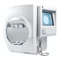
Zeiss HUMPHREY 750i User Manual (329 pages)
FIELD ANALYZER II - i series
Brand:
Zeiss
| Category:
Measuring Instruments
| Size: 20.13 MB
Table of Contents
Table of Contents
5
Introduction/Instrument Setup
9
About Visual Fields
10
What Visual Field Tests Measure
10
Normal Versus Pathologic Fields
11
The Normal Hill of Vision
11
The Boundaries of the Normal Visual Field
11
Methods of Testing the Visual Field
12
Patient Fixation and Test Reliability
12
The Humphrey Advantage
13
Reasons for Computerized Perimetry
13
Ergonomic Design
13
Easy Operation
14
Speedy Testing
14
Sophisticated Data Analysis with STATPAC™
14
Blue-Yellow (SWAP) Testing (Model 745I and 750I)
15
Automatic Fixation Monitoring
15
Data Protection Features
15
Information on the Internet
15
Using this Guide
16
Model Differentiation
16
Text Conventions
16
Additional References
16
Safety Precautions
17
General Safety Requirements
17
Installation Safety Precautions
17
Peripheral Placement Instructions
18
Radio and TV Interference
18
Symbol Definitions
19
Power on
19
HFA II Symbol Definitions
19
System Components
20
The HFA II – Side View
20
The HFA II – Rear View
20
The HFA II – Front View with Instrument Table
21
Additional Components
22
Printers
22
External Keyboard
22
Trackball, Mouse, or Other Input Device
23
External VGA Monitor
23
Surge Protectors
23
Connection of External Devices
23
Rear View of the HFA II with Panel Removed
24
Enlarged View of Cable Connections Panel on Rear of HFA II
24
System Assembly
25
The HFA II – Rear View on Instrument Table
25
General Operation
27
Screen Simplicity
28
General Information
28
The Title Bar
28
The Screen Body
28
Icon Buttons
29
The Information “I” Button
30
Touch Screen
30
The Unit Configuration Screen
30
Pop-Up Windows
31
Drop-Down Menus
31
Using the External Keyboard
32
Using the Trackball, Mouse, or Other Input Device
33
File Directory
34
Screen Saver
34
The Main Menu Screen
35
Command Buttons
35
System Setup
36
The Main System Setup Screen
36
Accessing the System Setup Screen
38
Altering the Language
38
Accessing Head Tracking (Model 750I)
39
Accessing the Vertex Monitor (Model 750I)
39
Setting the Time and Date
40
Printing the System Log
40
Accessing the Save/Transmit Option - Serial Connection
41
Accessing the Save/Transmit Option - Network Connection
42
Selecting the Printer Type
42
Selecting a Visual Acuity Format
43
Selecting Auto Pupil (Model 750I Only)
43
Personalizing Hard Copy Printouts
43
Altering the Main Menu Screen
44
Deleting a Button
45
Additional Setup
47
Adding Text to an Existing Button
47
Help Screens
49
Help Topics Screen
49
Setting-Up Tests
51
Selecting the Test Pattern and Test Eye
52
Using Test Buttons
52
Selecting Tests from the Main Menu Screen
52
Using the Test Library
53
Test Library
54
The Screening Test Library
55
The Threshold Test Library
56
The Specialty Test Library
56
Test Library Notes
57
Esterman Functional Tests
57
Entering Patient Data
58
Inputting Patient ID, Patient Name, Date of Birth, & Comments
58
Inputting Trial Lens Data
60
Clearing Patient Data
63
Recalling Patient Data
63
Patient Data 2 Screen
65
Inputting Diagnostic and Procedure Codes
65
Inputting Pupil Diameter and Visual Acuity
66
Inputting Intraocular Pressure (IOP)
67
Entering Cup/Disk (C/D) Ratios
68
Using Trial Lenses
69
Spherical Trial Lens Correction for Central Visual Field Testing
69
Inserting Trial Lenses into the Holder
71
Preparing the Patient
72
Patient Instructions for Static Testing
72
Occluding the Non-Test Eye
72
Seating the Patient
72
Positioning the Patient at the Instrument
73
No Trial Lenses for Esterman Monocular/Binocular Tests
74
Test Parameters & Strategies
75
Setting Test Parameters
76
To Change Test Parameters
76
The Screening Test Parameter Setup Screen
76
Standard Parameters
77
Test Strategies
78
Screening Test Parameters
78
The Threshold Parameter Setup Screen
81
Threshold Test Parameters
81
SITA ™ Testing
84
Tests Available with SITA
84
File Directory Indication
84
Floppy Disk Storage
84
Blue-Yellow (SWAP) Testing
85
Advantages of Testing with Blue-Yellow Perimetry
85
How Blue-Yellow Perimetry Works
85
Established Standards for Blue-Yellow Testing
85
Patient Selection for Blue-Yellow Perimetry
86
Blue-Yellow Testing
86
Extending the Visor Away from the Patient
87
Returning the Visor to the Retracted Position
88
Specificity of Blue-Yellow Testing
89
Alternate Color Testing (Models 740I -750I)
90
Performing a Color Test
90
Testing
91
Start Test Options
92
The Start of Test Screen
92
Monitoring and Maintaining the Patient's Eye Position
94
Video Eye Monitor
94
Gaze Tracking (Model 740I - 750I)
94
The Gaze Graph
95
Head Tracking (Model 750I)
95
Example of a Gaze Graph: Good Fixation with a Large Number of Blinks
95
Example of a Gaze Graph Indicating Poor Fixation
95
Vertex Monitor (Model 750I)
96
Supplemental Testing
97
Foveal Threshold
97
Gaze Tracking Initialization
98
Test in Progress
100
Printing Partial Tests
101
Fixation Monitoring
101
Tips for Gaze Tracking and Head Tracking
103
Test Complete Options
104
Saving to Disk
104
The Test Complete Screen
104
Do You Wish to Save this Test?
104
The (Threshold) Printout Selection Screen
105
Testing: a Step-By-Step Guide
106
Esterman Testing
108
Test Reliability
109
Factors Affecting Reliability
110
Patient Compliance
110
Patient Fixation
111
Trial Lenses
111
Evaluating Reliability
112
Fixation Losses
112
False Positive Errors
112
Sample Printout Showing Poor Reliability
113
False Negative Errors
114
Fluctuation Values
114
SITA Printout Showing Good Reliability
115
STATPAC Analysis & Printing Results
117
Introduction to STATPAC Analysis
118
Performing a STATPAC Analysis
118
STATPAC Threshold Formats
118
STATPAC Analysis & Printing Results
118
STATPAC Test Parameters
119
STATPAC Parameters for White-On-White Perimetry
119
STATPAC Parameters for Blue-Yellow Perimetry
119
Threshold Test Printout Formats
120
Reliability Indices
120
The Single Field Analysis Printout
121
The Glaucoma Hemifield Test
122
Superior Field Zones Used in the Glaucoma Hemifield Test
122
Total Deviation Plots
123
Pattern Deviation Plots
123
Global Indices
123
The Overview Printout
124
The Change Analysis Printout
126
The Box Plot
126
Change Analysis Summary of Global Indices
128
Linear Regression
128
A Mixed Change Analysis Printout with SITA Tests Included
130
The Glaucoma Change Probability Analysis Printout
131
The Change Probability Map
132
Change in Mean Deviation
132
The Glaucoma Change Probability - Baseline
133
The Glaucoma Change Probability - Follow up
134
Modified Linear Regression Analysis
135
On Statistics and Probability
135
A Note of Caution
135
Three-In-One Printout
136
SITA Printout Formats
137
An Example of the Overview Printout Showing both SITA Standard and SITA Fast Test Results for the same Patient
137
An Example of a Single Field Analysis Printout for SITA
138
Blue-Yellow Printout Formats
139
Single Field Analysis Printout
139
The Single Field Analysis Printout for Blue-Yellow (SWAP)
139
Glaucoma Hemifield Test
140
Overview Printout
140
The Overview Printout for Blue-Yellow (SWAP) Perimetry
140
Printing Current Threshold Test Results
141
To Print a Single Field Analysis of Current Test Results
141
To Print an Overview, Change Analysis, or Glaucoma Change Probability of Current Test Results
141
Screening Printout Formats
142
Reading Screening Test Printouts
142
Screening Printout Formats: All Screening Test Patterns
142
Printing Current Screening Test Results
143
Screening Printout Showing Results for both Eyes (the O. U. Printout)
143
Printing Previously Saved Test Results
144
Printing Test Results Stored on Disk
144
Printing from Recall Last Test
145
Printing from View Test
145
Printing Delay
145
Grayscale Symbols
146
Remote Printer Access
146
The Grayscale Shades Found on HFA II Printouts and Their Numerical Equivalents in Apostilbs (ASB) and Decibels (Db)
146
File Functions
147
File Functions Menu
148
Retrieving the File Directory
150
Disk Options
150
Source
150
Destination
150
Directory Order
151
The File Directory – Sorted by Patient
151
The File Directory – Single Patient Test Display
151
Selecting Tests from the Directory
152
The File Directory – Sorted by Name
152
Merging Patient Files
156
Performing File Functions
157
The View Test Screen
157
Transferring Tests from the HFA I to the HFA II
162
To Transfer Tests from the HFA I to the HFA II - I Series Via Serial Cable
163
To Transfer Tests from an Older HFA II to the HFA II - I Series Via Serial Cable
164
Database Status
165
Organizing Patient Files
166
Database Management
167
Introduction to Database Management
168
Database Integrity Functions
168
Patient Database Protection Procedures
169
Database Protection
169
Maintaining Backup Floppy and Magneto-Optical Disks
169
To Back up Hard Drive Data Onto Floppy Disks
170
To Back up Hard Drive Data Onto Magneto-Optical Disks
170
Set of Backup Floppy Disks with Labels
170
Configuration Backup and Restore
171
To Backup Configurations
171
To Restore Configurations
174
How to Handle Database Failures
176
Pathways for Resolving a Hard Disk Database Failure – an Overview
177
Hard Drive Failure: FIX NOW Option
178
Hard Drive Failure: FLOPPY ONLY Option
179
Hard Drive Failure: REBUILD HARD DISK DATABASE
181
Restoring the Hard Disk Database
184
To Restore the Hard Disk Database from Magneto-Optical Disks
185
To Restore the Hard Disk Database from Backup Floppy Disks
185
Floppy Disk Failure: REBUILD FLOPPY DISK DATABASE
185
Merge Database
186
Cleanup Hard Disk Database
187
Practices with Multiple Humphrey Field Analyzers
188
Care and Handling of Removable Storage Media
188
Custom Testing
189
Creating Custom Tests
190
Getting to the Custom Test Screen
190
An Example of the X, y Coordinate System for the Right Eye
193
Adding Single Points
194
Adding Grid Points
196
Combining Grids and Single Points
198
An Example of a Point and Grid Combination
199
Erasing Grids or Single Points
200
Saving Custom Tests
200
Deleting Custom Tests
203
Performing Custom Tests
205
Printout Format
207
An Example of a Custom Threshold Printout
207
Kinetic Testing
209
Introduction to Kinetic Testing
210
Performing Manual Kinetic Perimetry
211
Setting Parameters
213
Changing the Visual Field Size
215
Peripheral Testing Limits
216
Presenting a Stimulus Along a Meridian
216
Peripheral 80˚ Testing Limits for Selected Meridians
216
Choosing Meridians by Cursor or Keypad
217
Selecting the Meridian by Using the Cursor
218
Selecting the Meridian by Using the Keypad
219
Erasing Entries
221
Repeating Points Along the same Meridian
221
Pre-Defined Kinetic Test Patterns
222
Running Automated Kinetic Tests
223
Pausing the Test
225
Saving the Test Results
226
Step by Step Mode
227
Retesting a Meridian
229
Social Security Administration Kinetic Disability Test
232
The SSA Disability Test Printout
234
Special Mapping
235
Scotoma Mapping
236
Blind Spot Map
239
Static Points
241
Custom Scan
243
End of Test
246
Viewing Kinetic Tests
248
Printing Kinetic Tests
251
A Sample Kinetic Full Field Printout
251
Sample Kinetic Central Field Printout
252
A Sample Numerical Values Printout
253
Printout Legend
254
Printing Sequence
254
Designing a Custom Kinetic Test Pattern
256
Order of Stimulus Presentation
256
The Nasal / Temporal Relationship for the Right Eye
256
Erasing a Meridian
262
Saving the Custom Test
262
Naming the Test Button
263
Deleting Existing Kinetic Tests
264
Creating the Aphakic (Size IV) SSA Disability Test
265
Final Verification
270
Deleting the Test
270
Care and Cleaning
271
General Use Principles
272
Cleaning the HFA II
272
Air Intake Filter
273
Floppy Disk Drive
273
Replacing Parts
274
Stimulus Projection Bulb
274
Background Illumination Bulbs
276
Patient Response Button
276
Instrument Fuses
276
Power Table Fuses
278
HFA II Power Table with Mounted Printer Showing Location of Fuses
278
Operating the Printrex Printer
280
Loading Paper
280
Controls and Indicators
281
About Thermal Paper
281
Touch Screen Calibration
282
Using Data Disks
283
HFA II - I Series Product Specifications
285
Testing Features
286
Data Analysis Software
287
Printour Formats
287
Data Storage
287
File Sorting
287
User Features
288
H U M P H R E y F I E L D a N a L y Z E R
289
Icon Glossary
293
Goldmann Conversion Tables
295
Conversion of Goldmann Units to Decibels and Apostilbs
296
Conversion Tables: Goldmann Units to Decibels
297
Conversion Table for Blue-Yellow
298
Blue-Yellow Specifications
298
References to Section 4: Blue-Yellow (SWAP) Perimetry
298
Test Patterns
299
Threshold Test Patterns
299
Screening Test Patterns
301
Specialty Tests Patterns
304
Installing New HFA II Software
305
Software Installation
306
Additional Software Installation
307
How SITA Works / Acknowledgments
309
SITA Asks Smart Questions
309
SITA Tailors the Testing Pace to the Individual
309
SITA Knows When to Quit
310
SITA Carefully Recalculates All Threshold Values at the End of Testing
310
SITA Acknowledgments
311
Troubleshooting
313
Start-Up Difficulties
313
Time and Date
314
Printer
314
Printout Problems
315
Blind Spot Monitor
316
Gaze Tracking
316
Head Tracking
316
Vertex Monitoring
316
Pupil Size
317
Trackball / Mouse
317
Blue-Yellow (SWAP)
317
Testing Problems
318
Hard Disk Problems
318
Floppy Disk Problems
318
Magneto-Optical Disk Problems
318
Parts List
319
Index
321
Advertisement
Advertisement
Related Products
Zeiss HUMPHREY 720i
Zeiss HUMPHREY 745i
Zeiss HUMPHREY 740i
Zeiss humphrey HFA II-i series
Zeiss Humphrey II-i series
Zeiss Humphrey Field Analyzer 3
Zeiss VICTORY 7x42 T* FL
Zeiss Dialyt 7x50 B/GA T Marine
Zeiss opmi lumera 700
Zeiss Axioscope 7
Zeiss Categories
Microscope
Medical Equipment
Binoculars
Laboratory Equipment
Measuring Instruments
More Zeiss Manuals
Login
Sign In
OR
Sign in with Facebook
Sign in with Google
Upload manual
Upload from disk
Upload from URL