Sign In
Upload
Manuals
Brands
THORLABS Manuals
Laboratory Equipment
GANYMEDE
THORLABS GANYMEDE Manuals
Manuals and User Guides for THORLABS GANYMEDE. We have
1
THORLABS GANYMEDE manual available for free PDF download: Operating Manual
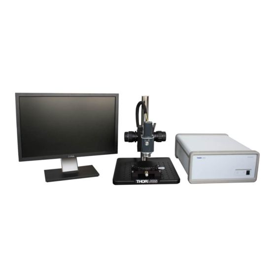
THORLABS GANYMEDE Operating Manual (60 pages)
Spectral Domain OCT System
Brand:
THORLABS
| Category:
Laboratory Equipment
| Size: 4 MB
Table of Contents
Table of Contents
2
Part 1. Introduction
7
Safety
8
Care and Maintenance
9
Optical Cleaning
9
Service
10
Accessories and Customization
10
Part 2. Description
11
Theory
11
Spectral Domain OCT Technology
11
Figure 1 Schematic Diagram of the SD-OCT System
11
Nomenclature in OCT Imaging
12
Figure 2 A-Scan Data Set
12
Figure 3 B-Scan Data Set
12
Figure 4 Rendered Volumetric Data Set
13
Figure 5 En-Face View or C-Scan
13
System Components
14
Packing List
14
Base Unit
16
Figure 6 Base Unit
16
Imaging Probe
17
Figure 7 Imaging Probe (Front View)
17
Figure 8 Imaging Probe (Rear View)
17
Figure 9 Optical Layout of Imaging Probe
18
Figure 10 Reference Intensity Adjustment
18
Figure 11 Reference Length Adjustment
19
Probe Stand
20
Figure 12 Probe Stand with Probe
20
Figure 13 OCT Stand Coarse Adjustment and Cable Clip(Rear View)
21
Figure 14 OCT Stand Adjusters and Dove Tail Slide
21
Figure 15 OCT Stand Sample Station
22
Graphical User Interface
23
Sdk
23
Warning Text
24
Figure 16 Laser Emission Warning Labels
24
Figure 17 Base Unit Warning Labels
24
Figure 18 Imaging Probe Warning Label
24
Part 3. Installation
25
Unpacking
25
System Connections
25
Figure 19 Rear View of Base Unit
25
Figure 20 Top View of the Probe with Interconnections
26
Figure 21 Gige Ethernet Adapter for Connection to Base Unit
26
System Installation
27
Figure 22 Mounting the Probe into the Dove Tail Slide of the OCT Stand
27
Figure 23 Cameralink Frame Grabber Card Inside the PC
28
Figure 24 Gige Cable Connected to Base Unit (A) and PC (B)
28
Figure 25
28
Figure 26 Plugging the Probe Connector into the Probe
29
Figure 27 Probe Connector Plugged into Probe
29
Figure 28 Installation of the Fiber at the Probe Fiber Connector
30
Figure 29 Installing the Probe Connection at the Base Unit
31
Figure 30 Installation of the Fiber at the Base Unit Connector
32
Figure 31 Protective Cap Removal
33
Part 4. System Operation
34
Starting the System
34
Basic Adjustments
34
Adjusting the Focus
34
Figure 32 Basic Focus Adjustment
34
Adjusting the Reference Intensity
35
Adjusting the Reference Length
35
Shutting down the System
36
Figure 33 B-Scan of an IR Viewing Card
36
Example Images
37
Figure 34 B-Scan of a Nailfold
37
Figure 35 B-Scan of a Fingertip
37
Figure 36 B-Scan of a Semi-Transparent Molded Plastic Cap
38
Figure 37 B-Scan of a Laminated IR Card (Not Included)
38
Figure 38 B-Scan of a Section of a Grape
38
Part 5. Imaging Artifacts
39
Saturation and Non-Linearity
39
Figure 39 High Surface Reflection Causing Saturation and Nonlinear Response of the Spectrometer
39
Wrong Reference Intensity Setting
40
Figure 40 Avoiding Strong Surface Reflection by Use of an Immersed Wedge
40
Figure 41 OCT Images Acquired with Low (A), High (B) and Good (C) Reference Intensity Setting
40
Auto-Interference
41
Figure 42 Auto-Interference from a Highly Reflective Sample in a B-Scan
41
Figure 43 Auto-Interference with the Reference Arm of the Imaging Probe Blocked
41
Multiple Scattering
42
Figure 44 Schematic of a Setup to Show the Influence of Multiple Scattering
42
Figure 45 OCT Image Showing Multiple Scattering
42
Phase Wrapping and Fringe Washout
43
Figure 46 Sample Image Showing Fringe Washout and Phase Wrapping
43
Flipped Image
44
Figure 47 Correct Reference Length Adjustment for Imaging of an IR Card
44
Figure 48 Incorrect Reference Length Adjustment Showing a Flipped Image
44
Shadowing
45
Figure 49 Rendered Volume of a Screw on an IR Viewing Card Displaying the Shadowing Effect
45
Image Distortion by Refractive Media
46
Figure 50 Schematic of a Setup to Show Distortions from Refractive Media
46
Figure 51 Height Shift of OCT Imaging through Refractive Media
46
The Group Refraction Index
47
Figure 52 Measurement Depth with Refractive Media
48
Measurement Depth in OCT Systems
48
Distortions in the Image
49
Figure 53 Different Materials in One Measurement
49
Figure 54 Complex Structure in Image
50
Part 6. Troubleshooting
51
Changing the Input Fuses
52
Figure 55 Fuse Cover on Base Unit Rear Panel
52
Part 7. Certifications and Compliance
53
Part 8. Warranty
54
Lasers and Imaging Systems
54
Non-Warranty Repairs
54
Warranty Exclusions
54
Part 9. Specifications
55
Part 10. Mechanical Drawings
56
Figure 56 Base Unit Dimensions
56
Figure 57 Imaging Probe Dimensions
56
Figure 58 Dimension of the Rotation Stage Underneath the Sample Plate
57
Part 11. Regulatory
58
Waste Treatment Is Your Own Responsibility
58
Ecological Background
58
Part 12. Thorlabs Worldwide Contacts
59
Advertisement
Advertisement
Related Products
THORLABS Ganymede Series
THORLABS GANYMEDE II HR
THORLABS GAN610
THORLABS GAN620
THORLABS GAN1x1 Series
THORLABS GAN1x1C1
THORLABS GAN3x1 Series
THORLABS GAN3x1C1
THORLABS GAN6x1 Series
THORLABS GAN6x1C1
THORLABS Categories
Measuring Instruments
Controller
Laboratory Equipment
Security Sensors
Industrial Equipment
More THORLABS Manuals
Login
Sign In
OR
Sign in with Facebook
Sign in with Google
Upload manual
Upload from disk
Upload from URL