Mindray Imagyn I9 Manuals
Manuals and User Guides for Mindray Imagyn I9. We have 3 Mindray Imagyn I9 manuals available for free PDF download: Operator's Manual

Mindray Imagyn I9 Operator's Manual (546 pages)
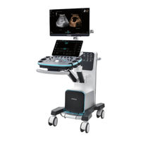
Diagnostic Ultrasound System
Brand: Mindray
|
Category: Medical Equipment
|
Size: 45 MB
Table of Contents
-
Warranty3
-
Latex Alert28
-
Intended Use29
-
Power Supply30
-
Options32
-
I/O Panel42
-
Keyboard47
-
Dialog Box50
-
Touch Screen51
-
Symbols54
-
Power ON/OFF60
-
Standby62
-
Ivocal71
-
Setup75
-
Region76
-
General77
-
Image Preset78
-
Application79
-
Key Board84
-
Gesture84
-
Output84
-
Workstation90
-
Biopsy90
-
Comment Preset100
-
List Config101
-
Iworks Preset102
-
View Management102
-
Protocol Edit103
-
Maintenance104
-
Dicom/Hl7104
-
Network Preset112
-
Network Settings112
-
Istorage Preset113
-
Medsight Preset113
-
Q-Path Preset114
-
MIOT Preset115
-
Print Preset115
-
Print Setting115
-
Image Settings115
-
Maintenance116
-
Load Factory116
-
Probe Check116
-
Other Settings117
-
Security118
-
Anti-Virus118
-
Exam Preparation121
-
Activate an Exam125
-
Continue an Exam125
-
Pause an Exam125
-
End an Exam125
-
Imaging Mode127
-
Image Adjustment127
-
B Mode130
-
Image Quality131
-
Line Density133
-
Color Mode137
-
Smart Track140
-
Power Mode141
-
Power Gain142
-
Color Map142
-
Flow142
-
Basic Operations143
-
M Mode146
-
Display Format147
-
Free Xros M149
-
PW/CW Mode150
-
Quick Angle154
-
Tdi154
-
Iscape View159
-
Basic Procedures159
-
Image Review160
-
Image Zooming160
-
Cine Review161
-
R-Vqs162
-
Smart B-Line163
-
Overview165
-
View Selection170
-
Curve Display171
-
Bulleye172
-
Data Export172
-
Fusion Imaging173
-
Overview173
-
Basic Procedures180
-
Marks186
-
Freehand 3D192
-
Measuring195
-
Smart VTI196
-
Smart IVC197
-
Screen Display200
-
View Operation200
-
Insert201
-
Create201
-
Overview203
-
Terms203
-
ROI and VOI204
-
About the Probes205
-
Mpr205
-
Free View206
-
Wire Cage206
-
Note before Use206
-
Static 3D208
-
Render Mode214
-
Rotate the Image215
-
Image Saving217
-
Color 3D217
-
Procedures218
-
Image Review219
-
Smart 3D219
-
Ilive222
-
Lighting Mode223
-
Layout224
-
Reference Point225
-
Print Format225
-
Smart Volume226
-
Basic Procedure226
-
Result Display228
-
Trace Mode229
-
Ipage230
-
Reference Image231
-
Image Rotation233
-
Scv233
-
Basic Procedures234
-
Smart Planes CNS236
-
Smart ICV238
-
Other Operations239
-
Basic Procedures240
-
Save the Image241
-
Basic Procedures242
-
Quick Adjustment242
-
Smart Face243
-
Volume CEUS244
-
Smart Scene 3D244
-
Elastography247
-
Image Parameters247
-
Mass Measurement249
-
Cine Review249
-
Image Parameters250
-
Measuring252
-
Cine Review252
-
Image Parameters253
-
Fatty Liver Lab255
-
Usat256
-
Hri257
-
Image Parameters259
-
Frame Average259
-
Contrast Imaging261
-
Image Parameters265
-
Ecg274
-
ECG Triggering274
-
Triggering Mode275
-
ECG Review275
-
Respiratory Wave275
-
Pcg276
-
Stress Echo279
-
Review/Wms Mode282
-
Smart Pelvic285
-
4D Image Data286
-
Res Zoom289
-
Pan Zoom290
-
Spot Zoom290
-
Cine Review291
-
Image Compare294
-
Frame Compare294
-
Icompare295
-
Cine Saving295
-
Live Capture296
-
Measurement297
-
Comments300
-
Adding Comments301
-
Adding an Arrow301
-
Moving Comments302
-
Editing Comments302
-
Voice Comments303
-
Body Mark304
-
Adding Body Mark305
-
Dicom/Hl7307
-
DICOM Storage308
-
Encapsulate PDF309
-
Unload DCM File309
-
DICOM Print309
-
Worklist310
-
Mpps311
-
Query/Retrieve312
-
Media Storage313
-
Media Review313
-
Data Restore313
-
Storage Media315
-
Thumbnails317
-
Image Review317
-
Image Analysis318
-
Report Storage320
-
Recycle bin322
-
Istorage323
-
U-Link323
-
Print323
-
Image Print323
-
Report Printing324
-
Log Upload325
-
Task Status325
-
Q-Path325
-
V-Access326
-
Probes327
-
Biopsy Guide342
-
Disposal388
-
Installation391
-
Mark398
-
Disposal401
-
Middle Line401
-
DVR Recording403
-
Start Recording403
-
Send Image403
-
DVR Video Replay404
-
Replay on PC404
-
Checking Battery411
-
Troubleshooting413
-
Barcode Reader415
-
Setting416
-
Volume Setting417
-
Overview421
-
Setting422
-
Wireless LAN427
-
IP Configure428
-
EAP Network429
-
D Ivision434
-
Acoustic Output435
-
MI/TI Display437
-
Adjusting Range437
-
Acoustic Output438
-
Power Cord Plug441
-
Device Labeling442
-
Operation455
-
Structure459
-
Specifications459
-
Specification461
-
Interoperability464
-
Security Policy465
Advertisement
Mindray Imagyn I9 Operator's Manual (477 pages)
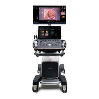
Diagnostic Ultrasound System
Brand: Mindray
|
Category: Medical Equipment
|
Size: 15 MB
Table of Contents
-
Warranty8
-
Conventions11
-
-
Imaging Mode75
-
Flow94
-
Free Xros M Mode104
-
Tdi106
-
Iscape View155
-
Contrast Imaging158
-
Elastography165
-
Stress Echo176
-
Fusion Imaging190
-
R-Vqs217
-
Smart VTI221
-
Smart IVC222
-
Smart B-Line224
-
-
-
Cine Review229
-
Image Compare231
-
Cine Saving233
-
-
Ecg236
-
Respiratory Wave237
-
-
8 Measurement
241 -
-
Comments247
-
Voice Comments251
-
Body Mark252
-
-
-
Istorage268
-
Print269
-
Administration271
-
Q-Path279
-
V-Access280
-
11 Dicom/Hl7
281 -
12 Setup
303-
System Preset303
-
Exam Mode Preset311
-
Comment Preset312
-
Iworks Preset313
-
DICOM/HL7 Preset313
-
Network Preset314
-
Print Preset317
-
Maintenance318
-
Security319
-
-
-
Probes321
-
Biopsy Guide339
-
Middle Line378
-
-
14 DVR Recording
381-
Send Image381
-
DVR Video Replay382
-
Mindray Imagyn I9 Operator's Manual (475 pages)
Diagnostic Ultrasound System
Brand: Mindray
|
Category: Medical Equipment
|
Size: 13 MB
Table of Contents
-
Warranty8
-
Conventions11
-
Latex Alert21
-
Intended Use23
-
Symbols40
-
Imaging Mode75
-
Flow94
-
Free Xros M Mode104
-
Tdi106
-
Iscape155
-
Contrast Imaging158
-
Elastography165
-
Stress Echo176
-
Fusion Imaging190
-
R-Vqs217
-
Smart VTI221
-
Smart IVC222
-
Smart B-Line224
-
Cine Review229
-
Image Compare231
-
Cine Saving233
-
Ecg236
-
Respiratory Wave237
-
Measurement241
-
Basic Operations241
-
Comments247
-
Voice Comments251
-
Body Mark252
-
Istorage268
-
Print269
-
Administration271
-
Q-Path279
-
V-Access280
-
Dicom/Hl7281
-
DICOM Preset281
-
DICOM Verifying296
-
DICOM Services296
-
Setup303
-
System Preset303
-
Exam Mode Preset311
-
Comment Preset312
-
Iworks Preset313
-
DICOM/HL7 Preset313
-
Network Preset314
-
Print Preset317
-
Maintenance318
-
Security319
-
Probes321
-
Biopsy Guide339
-
Middle Line378
-
DVR Recording381
-
Send Image381
-
DVR Video Replay382
-
Acoustic Output383
-
Acoustic Output386
Advertisement