Sign In
Upload
Manuals
Brands
Imaging Sciences Manuals
Medical Equipment
i-Cat 17-19
Imaging Sciences i-Cat 17-19 Manuals
Manuals and User Guides for Imaging Sciences i-Cat 17-19. We have
1
Imaging Sciences i-Cat 17-19 manual available for free PDF download: Operator's Manual
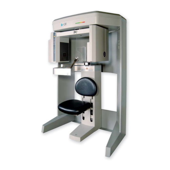
Imaging Sciences i-Cat 17-19 Operator's Manual (292 pages)
Cone Beam Volumetric Tomography and Panoramic Dental Imaging System
Brand:
Imaging Sciences
| Category:
Medical Equipment
| Size: 11.4 MB
Table of Contents
Table of Contents
3
What's New in this Release
11
Chapter 1 - Introduction
13
System Description
13
Intended Use of the Device
14
Major System Items
15
About the Operators' Manual
15
Conventions Used in the User Manual
15
Standard Limited Warranty
16
Backup Recommendations
17
Chapter 2 - Safety Items
19
Important Safety Information
19
Warnings, Cautions, and Notes
19
Safety Precautions
20
Electrical Hazards
20
Explosion Hazard
21
Mechanical Hazards
21
Laser Beam Hazards
22
Radiation Safety
23
Radiation Protection Measures
23
System Safety Devices
24
Emergency Stops
24
Warning System
24
Interlock System
24
Sample Site Plan
25
Interlock and Warning System Schematic
26
Cabling Requirements
27
Emergency Removal of a Patient
27
Error Messages
27
System Labels
28
Chapter 3 - System Controls and Indicators
37
Operator Control Box
37
Patient Emergency Stop Control
38
Patient Alignment Panel
38
System Status Indicators
38
Chapter 4 - System Startup and Shutdown
39
System Startup
39
System Shutdown
39
Chapter 5 - Managing Patient Data
41
Patient Information
41
Study Information
42
Hide/Display Study List
44
Delete Patient Scans
44
Add New Patient
45
Edit Patient Details
46
Access Patient Data
47
Delete a Patient
48
Chapter 6 - Patient Positioning and Protocols
49
Patient Positioning
49
Patient Chair
50
Gate
51
Chin Support
51
Head Support and Head Strap
52
Alignment Light
53
Instruct Patients Prior to Exam
55
Previews and Dry Runs
55
Protocol Guidelines
56
Chapter 7 - Acquisitioning (Scanning)
57
Volume Scans
57
Preview, Dry Run, and Capture Scans
59
Dose Area Product Feature
62
Quick Picks
64
PAN Scans (Optional)
65
Chapter 8 - Reconstruction of Anatomy
71
Preview Screen
71
Using Quality Control Frames
73
Panoramic View (Tru-Pan Feature Set as Default)
75
Manually Adjusting the Panoramic View
78
Removing Circumference Artifacts
80
Adjusting MIP, Centerline and Image Type
81
Selecting MIP or Radiograph Display
81
Adjusting Brightness and Contrast
82
Pan Feature
82
Rotation Feature
82
Zoom Feature
83
Back Tool
83
Filter Settings
83
Taking Measurements
85
Hounsfield Units
85
Distance
88
Rotating Views
90
Saving Views as JPEG Image Files
91
Saving and Loading Workups
91
Save a Workup
91
Load an Existing Workup
93
Delete an Existing Workup
94
Viewing and Reconstructing Raw Patient Scans
95
Quantum IQ
97
Chapter 9 - Detail Screens
99
Preview Screen
99
Implant Planning Screen
100
Estimate the Nerve Canal
105
Ceph Screen
114
MPR Screen
116
TMJ Screen
120
Chapter 10 - Tools
123
DICOM Setup
123
DICOM Database and DICOM Export Setup
123
Pacs
125
DICOM Character Set for New Files
125
Check Read/Write Access to Image Database
127
Export DICOM
128
Export an Original CT or PAN Study
128
Export a Rotated CT Study
129
Export Sure Smile Studies
129
Create Export CD
130
Erase CD-RW
132
Output to Folder
132
Import Study
133
Reporting
134
Run Report
134
Create New Report
135
Modify Existing Report
139
Start External Applications
139
Chapter 11 - Calibration
141
Panel Calibration
141
Collimators Calibration
143
Geometry Calibration
144
Chapter 12 - Quality Assurance
149
QA Phantom Test
149
Line Pair Evaluation
152
Distance Measurement Test
153
Hounsfield Unit (HU) Measurements
154
QA Water Phantom Test
157
Noise Level Test
160
Uniformity Test
161
PAN Phantom Test
162
Radiation Output Test
165
Measured Dose
166
Interpretation
166
Chapter 13 - Radiation Environment Survey
167
Conditions of Operation - 8.9 and 26.9 Second Scans
168
Scatter Measurements for 8.9 Second Scans
168
Diameter 16Cm 8.9 Second Scan
169
Scatter Measurements for 26.9 Second Scan
170
Diameter 16Cm 26.9 Second Scan
170
Conditions of Operation - PAN Scans
171
Scatter Measurements for 20 Second PAN Scan, Large 2X2
172
Scan Times and Settings
173
Dose and Imaging Performance Information
174
Dose Profile
175
Sensitivity Profile
177
Drywall Attenuation
178
Recommended Operating Requirements
179
X-Ray Tube Assembly
180
Chapter 14 - Product Information
185
Technical Specifications
185
Power Requirements
186
Apparent Resistance of Supply Mains
187
Weight
187
Environmental Specifications
188
Operating
188
Transportation and Storage
188
Acquisition Computer
188
Patient Support Chair
188
Disposal
188
Extension Cords
189
External Item
189
Cleaning
189
Electromagnetic or Other Interference (Emissions and Immunity)
189
Equipment Standards
191
Equipment Class
191
Preventive Maintenance Schedule - for Owner / User
192
Planned Maintenance - 12 Month Schedule
192
Cleaning
193
Planned Maintenance Checklist
193
Replaceable Parts
194
Supplemental Components
195
Optional PAN Scan Accessories
196
System Gantry Dimensions
197
Chapter 15 - Networking Support Setup
199
Networking Data Flow
199
Networking Support Overview
199
Network Support Installation/Setup
200
Provide Network Storage
200
Install Sweeper Service
201
Configure Sweeper Service
201
Migrate from Standalone to Server
202
Change Image Root Folder
203
Start the Sweeper
203
Sweeper Service
203
Sweeper Controller
204
Network Failures
204
No Network Access at Startup
205
Network Fails During Operations
206
Limitations of Networking Support
206
Chapter 16 - Remote System Import and Export
207
Remote System Import and Export Overview
207
Remote System Import
207
Import Installation and Setup
208
Set up DICOM Worklist Interface
208
Set up Practice Management Interface
210
Import Patient Data
211
Remote System Export (RSSM)
212
RSSM Installation and Setup
212
RSSM Logs
216
Send Patient Data to a Remote System
216
Rsqm
218
RSQM Installation and Setup
218
Query and Retrieve Images
221
Status Messages
222
Appendix A - Icatvision & Icattransfer
225
Workstation Minimum Requirements
226
Laptop Minimum Requirements
226
File Structure Setup
226
Install Icatvision
227
Icatvision Setup
228
Icattransfer
228
Install Icattransfer
229
Icattransfer Passcode
229
Icattransfer Setup
230
Configure Icattransfer
230
Accessing I-CAT DICOM Data
231
Appendix B - Three Dimensional Volume Rendering (3DVR)
233
Open Database
233
Axial Functions
234
Paging
234
W/L (Window/Level
235
ROI (Region of Interest
235
Distance
236
Identify
236
Remove Object
238
Hounsfield Unit Calibration Offset
238
View Volume
240
Projection Type
241
Volume Edit
242
VR Functions
243
Pop up Menu
246
View Full Screen
247
Appendix C - Quick Reference
249
Navigating the Interface
249
Tools for Viewing this Image
249
Suggestions for Adjusting Panoramic Map
250
Filtering Defaults
250
Removing Circumference Artifact
250
Saving and Loading Workups
250
Keyboard Shortcuts
250
Implant Planning Screen
251
Panoramic Map
252
Axial Slice Position
252
Ceph Screen
253
MPR Screen
253
Install Case Studies from Cds
254
TMJ Planning Screen
254
Create Export Cds
254
Appendix D - System Labels
255
English
255
Français
257
Deutsch
259
Italiano
261
Polski
263
Portuguê
265
Español
267
Svenska
269
简体中文 (Chinese
271
繁體中文 (Taiwan
273
한국어 (Korean
275
日本語 (Japanese
277
Česky
279
Nederlands
281
Русский Язык
283
Român
286
Türkçe
288
Advertisement
Advertisement
Related Products
Imaging Sciences Categories
Medical Equipment
More Imaging Sciences Manuals
Login
Sign In
OR
Sign in with Facebook
Sign in with Google
Upload manual
Upload from disk
Upload from URL