Table of Contents
Advertisement
Advertisement
Table of Contents

Related Manuals for YHLO BIOTECH VISION ESR

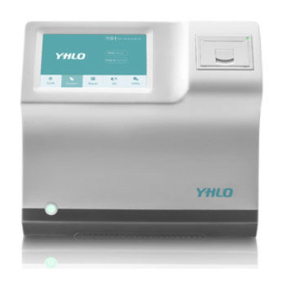
Summary of Contents for YHLO BIOTECH VISION ESR
- Page 1 VISION ESR Analyzer Operation Manual Released Software V1...
- Page 3 ©2016-2021 SHENZHEN YHLO BIOTECH CO., LTD. All rights Reserved. This Operation Manual is released in 01 February 2020. Customer service department Manufacturer: SHENZHEN YHLO BIOTECH CO., LTD. Address: Building 1, YHLO Biopark, Baolong 2nd Road, Baolong Subdistrict, Longgang District, 518116 Shenzhen, PEOPLE’S...
- Page 4 Contents of this manual are subject to change without prior notice. All information contained in this manual is believed to be correct. SHENZHEN YHLO BIOTECH CO., LTD. (hereinafter called YHLO) shall not be liable for errors contained herein or for incidental or consequential damages in connection with the furnishing, performance, or use of this manual.
- Page 5 Warranty This warranty is exclusive and is in lieu of all other warranties, expressed or implied, including warranties of merchantability or fitness for any particular purpose. Exemptions YHLO's obligation or liability under this warranty does not include any transportation or other charges or liability for direct, indirect or consequential damages or delay resulting from the improper use or application of the product or the use of parts or accessories not approved by YHLO or repair by people other than YHLO authorized personnel.
- Page 6 Intellectual Property Statement SHENZHEN YHLO BIOTECH CO., LTD. owns the intellectual property rights to this YHLO product and this manual. This manual may refer to information protected by copyright or patents and does not convey any license under the patent rights or copyright of YHLO, or of others.
- Page 7 Restriction and Warning Please use the components provided or recommended by the manufacturer (YHLO). Any risk or malfunctions resulted from disobey of the manufacturer’s operation manual should be assumed by the users. The running of the instrument is dependent on the corresponding software. Any damage and hacking towards the software should be considered as the infringement of the guarantee agreement.
-
Page 9: Table Of Contents
Table of Contents Introduction ............................ 1 Field of Application ....................... 1 Instrument Structure ......................1 Operation Accessories ......................2 Technical Data ........................2 Installation Requirement ...................... 3 Safety Instruction .......................... 4 Operator..........................4 Tube Requirement ........................ 4 Sample ..........................4 Symbol .......................... - Page 10 Test Procedures in “Random” Mode ................... 28 Service and Maintenance......................29 Troubleshooting .......................... 29 Fault Content ........................29 Fault Causes and Handling ....................30 Quick Guide......................... 1 Appendix Precautions for Operation ..................3 Appendix Sample Labeling ......................4 Appendix Sedimentation Rate Correction for Variations in Room Temperature ....5 Appendix Sedimentation Rate Correction for Anemia Sample ..........
-
Page 11: Introduction
1. Introduction 1.1 Field of Application VISION Pro (hereinafter referred to as the instrument) is a clinical diagnostic instrument for determining the sedimentation value of blood sample. Used together with disposable EDTA tubes, the sample positions are 8, 16 and 32. During the test, the sample tube rack will be rotated 180°... -
Page 12: Operation Accessories
1.3 Operation Accessories The accessories are included in the package list, please see the attachment for details and confirm. If any of the mentioned parts is lost or damaged, please contact YHLO technicians or local distributors. 1.4 Technical Data Display 7 inch LCD touch Screen Power Indicator LED, Green... -
Page 13: Installation Requirement
1.5 Installation Requirement Temperature Range Moisture 40%RH 80%RH Atmospheric Pressure 75.0kPa 106.0 k Pa Power Supply 100-240V~, 50/60Hz instrument should mounted well-ventilated room and connected with a three-wire power cord good grounding Miscellaneous performance. Avoid strong magnetic disturbance, keep away from explosion gas, dust, direct light and water immersion. -
Page 14: Safety Instruction
2. Safety Instruction 2.1 Operator The instrument is exclusively used for clinical diagnostics. The operator is only limited to the YHLO engineers and personnel trained by the dealers. 2.2 Tube Requirement Any EDTA plastic or glass tubes within a diameter of 12 to 13mm are applicable. The scanner can scan the sample through the trademark and barcode labels attached to the tube. -
Page 15: Symbol
2.4 Symbol You may find the following symbols on the analyzer. Symbols Meaning Attention! This symbol identifies a safety note. Ensure you understand the function before you operate. WARNING. Biological hazard. WARNING. Laser beam WARNING. Be careful with hand. Ethernet port USB port SD card slot Ordering code... -
Page 16: Instrument Identification
2.5 Instrument Identification Information listed on Identification Plate: Name of Instrument Model In vitro Diagnostics Instrument Marking Electrical Requirements and Maximum power consumption Serial Number Date of Manufacturing CE mark Registration Number Name of Manufacturer Address of Manufacturer 2.6 Emergency Power Switch In case of emergency, cut off the electrical supply to the instrument by turning off the power switch located at the back of the instrument, or unplug the power outlet (The instrument should be placed in which the power switch is easy to access). -
Page 17: Instrument Installation
3. Instrument Installation 3.1 Preparation 3.1.1 Installation Personnel The installation personnel are only limited to YHLO engineer or personnel authorized by customer service supplier. 3.1.2 Delivery, Storage and Unpacking Fig. 3-1 Package Indicator Please following the below requirements during delivery and storage. Do not store the instrument in humid environment. -
Page 18: Installation
Check the instrument and components in accordance with the packing list when unpacking. Softly place the instrument on the workbench. Check the serial number on the instrument nameplate, conformity certificate and the delivery list for consistency. Scrutinize the instrument to check if there are loose, bend or cracked components. -
Page 19: Software Operation Instruction
4. Software Operation Instruction 4.1 System Start Connect the power cord. Turn on the power switch on its back. The Login interface of the instrument displays. Type in the user name and password, click “Login” to enter the main interface Fig. - Page 20 Serial Symbol icons Meaning It shows the LOGO of manufacturer. It shows Instrument status. It shows current instrument temperature, real-time display update. It shows LIS connection status. It shows printer connection status. It shows the instrument state, and can be clicked into the failure detail list.
-
Page 21: Menu Tree
4.2 Menu tree The following is a listing of the VISION Pro menu tree. Cycle Test Interface 4.2.1 Click “Cycle” and the instrument can perform a self check to make sure the instrument connection, lifting and mixing motors normal functions, home positions, infrared LED. - Page 22 Attention The "Previous" and "Next" only display in the VISION Pro-B or VISION Pro-C. Cycle: Indicates the current function interface. Home: Click "Home" to back the main interface. Fig. 4-2-1-2 Edit Sample Info Interface Position: The current sample position will be placed. Sample ID: Shows the current Sample ID.
-
Page 23: Random Interface
Fig. 4-2-1-4 Edit Sample Info Interface Save: Save the patient information. Previous: Goes to the previous position. Next: Goes to the next position. Back: Back to the Cycle interface. Patient ID: Enter the patient ID. Sample ID: Shows the current Sample ID. Name: Enter the name of patient. - Page 24 Fig. 4-2-2-1 Random Interface Edit: Click “Edit” to enter the interface shown in Fig. 4-2-2-2 and Fig. 4-2-1-3 below. >>/<<: Click ">>" to go to the previous page, click "<<" to go to the next page. Fig. 4-2-2-2 Edit Sample Info Interface Position: The current sample position will be placed.
-
Page 25: Quality Control
Next: Goes to the next position. Back: Back to the Cycle interface. The description of patient information is the same as the "Cycle" ( Fig. 4-2-1-3). Quality Control 4.2.3 Quality control testing will use 2 levels of quality control, a high level and a low level. - Page 26 Fig. 4-2-3-1 QC Interface Setting: Click "Setting", the interface appears two levels of quality control products C1, C2, input the quality control number, reference mean, deviation range respectively and save. The interface will appear the quality control product information list that has been entered. Fig.
- Page 27 Note The same batch quality controller need to have only one control information. L-J: L-J chart takes the test time as the horizontal axis, the test result as the longitudinal shaft. It shows the controller result tendency in the specified time period.
- Page 28 Turn on the power on the back of the instrument .After the boot interface is finished, input the user and password, click "Login". Click "Cycle", then click "Scan ID" to begin the scan the self-check process will be run automatically. If the self-check goes through, the link will show green color.
-
Page 29: Result Interface
Result Interface 4.2.4 Fig. 4-2-4-1 Result Interface Select “Result” in the main interface to view the test results. The “Position” column in black shows whether tests were performed in “Cycle” mode or “Random” mode. The “ESR” and “ESR (18°C)” columns both indicate results. The “ESR”... - Page 30 Fig. 4-2-4-2 Search Interface Fig. 4-2-4-3 Curve Interface V3.0 OM- VISION Pro valid from 01 February 2020...
-
Page 31: Settings Interface
Settings Interface 4.2.5 Fig. 4-2-5-1 Settings Interface 4.2.5.1 Sys Set Interface Fig. 4-2-5-1 and Click “Settings” in the main menu to enter the display shown above (See Fig. 4-2-5-5 Instrument- Language: Select the corresponding language. After a restart, the language will be changed. Instrument- Settings: Select “Auto Print”... - Page 32 Fig. 4-2-5-3 LIS Settings Interface Instrument-Version: In the picture below(See Fig. 4-2-4-4), you can know the version of software. Fig. 4-2-5-4 Sys Set-Version Interface 4.2.5.2 Aux Interface Fig. 4-2-5-5 The interface is used to set the reference, user, log out and dictionary. (See V3.0 OM- VISION Pro valid from 01 February 2020...
- Page 33 Fig. 4-2-5-5 Aux Interface Aux-Reference: This menu allows user to set the reference range of results. Add: Click “Add” to add this reference range. Delete: Select a reference range to be deleted on the left side, then click "Delete" to delete this reference range. Modify: Select a reference range to be modified on the left side;...
- Page 34 password or permission. When you finished the modification, click the "Modify". Delete: Elect the user that you want to delete, click the "Delete". Aux-Log out: This menu allows user to back to login interface. Fig. 4-2-5-7 Aux-Log out Interface Aux-Dictionary: This menu allows user to add or delete the classification. Fig.
-
Page 35: Instrument Operation
5. Instrument Operation The instrument is required to run a self-check before analysis. There are two test modes for the instrument, “Cycle” and “Random”. Press “Cycle” to perform tests and the sample will be mixed automatically. Press “Random” to perform tests and the sample should be mixed by hand before test. During the test, new samples can be inserted into the instrument. -
Page 36: Flow Chart Of Operation
5.1 Flow chart of Operation Switch On Result Reporting Self Check Data Processing Sample Scanning Select sample position Barcode Scanning or Sample Mixing Input Sample ID Put sample tubes into the rack Start Running Fig. 5-1 Flow chart of operation (Cycle) Switch On Result Reporting Self Check... -
Page 37: Test Procedures In "Cycle" Mode
5.2 Test Procedures in “Cycle” Mode Connect the power cord. If the instrument is a limited license, please insert a dongle. Turn on the power switch on its back. 1) After the boot interface is finished, input the user and password, click "Login". 2) Click "Cycle", then click "Scan ID"... -
Page 38: Test Procedures In "Random" Mode
5.3 Test Procedures in “Random” Mode Connect the power cord. If instrument is a limited license, please insert a dongle card. Turn on the power switch on its back. 1) After the boot interface is finished, input the user and password, click "Login", then click "Random"... -
Page 39: Service And Maintenance
6. Service and Maintenance VISION Pro employs an enclosed design and will not produce waste liquid during test. The instrument will not come into contact with blood samples, and it does not contain other parts requiring routine maintenance. Only the following maintenance will be carried out when the device is used normally: Remove dust on the surface of the device, including the light transmitting window of scanner. -
Page 40: Fault Causes And Handling
Fault Content Meanings Mixing motor home failed! Error when the mixing motor returns to home position Linear motor home failed! Error when the linear motor returns to home position Dark reading error : Channel Dark light reading error of XX channel Weak light reading error : Channel Weak light reading error of XX channel Strong light reading error : Channel... - Page 41 reversely connected; 5) Test whether the 24V supply voltage to the main board is normal; replace the abnormal power supply; 6) Replace the motor; 7) Replace the main board. Dark/weak/strong light reading error: Dark/weak/strong light reading error of channel Fault cause: 1) In a strong-light environment (e.g.
- Page 42 2) Move the optical component to the home position; enable the Weak/Strong button of the error reporting channel in sequence. If the LED reading of the error reporting channel is less than 3500 and the optical reading of the right-side channel is also much lower than the normal value (weak light: around 1500;...
-
Page 43: Appendix Quick Guide
Appendix Quick Guide 1 Switch On Check and ensure the power cord well connected. Switch on the Instrument by means of the power switch on its back. After self check, the software main menu will be displayed. 2 Preparation for Sample Analysis The sample can be used for testing within 4 hours after sampling and should always be kept in the room temperature. - Page 44 B: Perform test in “Random” mode Click "Random" to enter the interface. Open the lid of instrument, select the position, then click "Edit"; Click the input box of "Sample ID", use the inner barcode reader to scan the sample barcode or manually enter the sample barcode, select the "Reference"...
-
Page 45: Appendix Precautions For Operation
Appendix Precautions for Operation Operation Requirement: 1. Please run tests under 18 C ~30 C and keep the room temperature stable. 2. Do not run tests in vibrant or dusty environment. Sample Requirement: 1. Blood samples should be tested within 4 hours after collection. 2. - Page 46 Appendix Sample Labeling tube label barcode label sample tube receiver Highly recommended layer label Acceptable Highly recommended Acceptable layer labels V3.0 OM- VISION Pro valid from 01 February 2020...
- Page 47 Appendix Sedimentation Rate Correction for Variations in Room Temperature It is noted that sedimentation rates vary with room temperature. In high temperature, low red blood clumping cause sedimentation at faster rate; in low temperature, intensive red blood clumping result in slower sedimentation rate.
-
Page 48: Appendix Sedimentation Rate Correction For Anemia Sample
Appendix Sedimentation Rate Correction for Anemia Sample Anemia is a medical condition in which there are too few red cells in blood. Westergren’s Method is the international standard method made by ICSH to perform ESR test. When Westergren’s method is performed to test anemia samples, the test result is higher than the real sedimentation rate due to the reasons below: The sedimentation curve is often divided into three components: (1) an initial slow phase or period of... - Page 49 Observed Rate (Westergren’s Method) Correction Line Fig.A-2 Ordway-Singer Nomogram Sedimentation Rate Correction for Anemia Sample (Westergren’s Method V3.0 OM- VISION Pro valid from 01 February 2020...
Need help?
Do you have a question about the VISION ESR and is the answer not in the manual?
Questions and answers