Advertisement
Table of Contents
All cited trademarks are the property of their respective owners. CAUTION: The law restricts these devices to sale by or on
the order of a physician. Indications, contraindications, warnings and instructions for use can be found in the product labelling
supplied with each device. Information for the use only in countries with applicable health authority product registrations.
PSST 4893 Printed in Germany by medicalvision.
FV3_SpyglassGuide.indd 1-2
SpyGlass
Direct Visualisation System and Capital Components
www.bostonscientific.com
www.bostonscientific-international.com
RCS Nanterre B420 668 420
© 2008 Boston Scientific Corporation
or its affiliates. All rights reserved.
DINEND2174EA
™
User Reference Guide
19.05.2008 12:51:35 Uhr
Advertisement
Table of Contents

Related Manuals for Boston SpyGlass

Summary of Contents for Boston SpyGlass
- Page 1 Indications, contraindications, warnings and instructions for use can be found in the product labelling © 2008 Boston Scientific Corporation supplied with each device. Information for the use only in countries with applicable health authority product registrations.
-
Page 2: Table Of Contents
Intended Use This overview is provided for illustrative purposes only and is intended only as a brief summary of how the procedural steps for using the SpyGlass Direct Visualisation System are generally performed. Please refer to the Directions For Use ™... -
Page 3: Spyglass™ System Capital Components And Consumable Devices
Consumable Devices (Figure 1) The capital system contains several components that will require assembly: • SpyGlass Camera – Auto shutter camera with 6.4 mm CCD chip SpyGlass Ocular • SpyGlass Ocular – optical coupler that interfaces with the SpyGlass Probe... -
Page 4: Verifying Power
• Do not shine light directly into the camera as this may damage the imaging sensor. Figure 4 NOTE: A clear image will not be seen until the SpyGlass™ Probe is connected. FV3_SpyglassGuide.indd 4-5 19.05.2008 12:51:36 Uhr... -
Page 5: Pre-Procedure Setup
Pre-Procedure Set-Up The SpyGlass System procedure is an extension of a typical ERCP, so initial steps ™ are the same as or similar to an ERCP. Pre-procedure device set-up is required. A review of select pre-procedure set-up and typical procedural steps are listed below. -
Page 6: Image Quality Test
Turn the lightsource to a mid-level power NOTE: Lightsource cable should be attached to SpyGlass Probe at this point. • Verify a proper image using the SpyGlass Probe test target. Refer to the white index card included in the SpyGlass Probe packaging. •... -
Page 7: Preparing And Using The
Ensure the probe is pulled back into the catheter several millimeters to avoid damage to the probe. • Using short strokes, insert the SpyScope Catheter and SpyGlass Probe into the working channel of the duodenoscope, over the elevator and into the Ampulla of Vater. -
Page 8: Accessory Passage
Accessory Passage • If not already preloaded, insert the SpyBite Biopsy Forceps into the device port ™ of the SpyScope Catheter. ™ (Figure 10) • Advance the SpyBite Biopsy Forceps using short strokes. Once the forceps exit the tip of the SpyScope Catheter, the forceps can be extended to obtain a tissue sample. -
Page 9: Probe And Ocular Care And Storage
• With proper handling and care, the SpyGlass Probe can be used for multiple uses. Before each use, the probe is reprocessed according to the recommended methods identified in the reprocessing section of the SpyGlass Probe Directions for Use. -
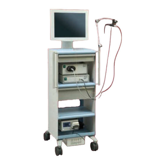
Page 10: Spyglass System Components
M00546451 SpyGlass Irrigation Tube Set (Box 10) NOTE: SpyGlass System, when completely assembled, has an approximate space footprint of 49.5 cm W x 53.3 cm D. Cart includes a 3-joint arm with connector for ocular, but no table clamp Total weight of the assembled system is approximately 97.068 kg. -
Page 11: Intended Use
SpyGlass Direct Visualisation Probe and Ocular SpyGlass Lightsource INTENDED USE The SpyGlass Probe and Ocular have been designed to examine the biliary system and associated ducts and organs. INDICATIONS FOR USE Boston Scientific SpyGlass Lightsource is used for surgical lighting and other applications. - Page 12 Notes FV3_SpyglassGuide.indd 20-21 19.05.2008 12:51:38 Uhr...
Need help?
Do you have a question about the SpyGlass and is the answer not in the manual?
Questions and answers