Table of Contents
Advertisement
Quick Links
Download this manual
See also:
User Manual
Advertisement
Table of Contents

Related Manuals for Zeiss AxioObserver


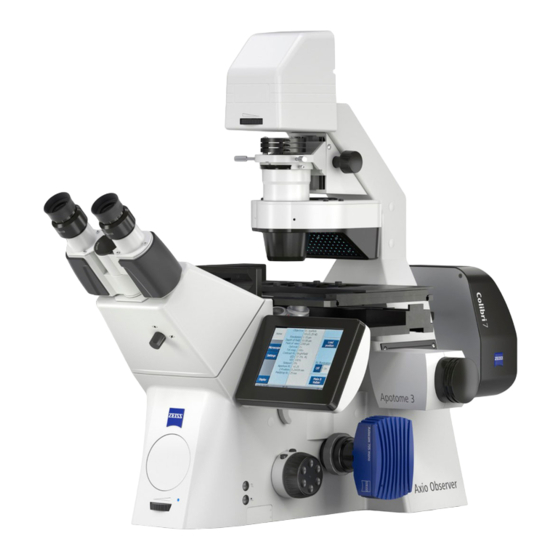
Summary of Contents for Zeiss AxioObserver
- Page 1 Zeiss AxioObserver with ApoTome Quick Start User Guide (AxioVision4.8) LSU Health Sciences Center-Shreveport Research Core Facility (RCF) Microscopy...
-
Page 2: Table Of Contents
RCF Microscopy Manual 5/2/2017 Authors Cory Nook and Chaowei Shang Table of Contents Start up the system…………………………………………………………………………………………………….. Page Touch screen controller (TFT DISPLAY) Page Mount and view the sample through the microscope ………………………………………………. Page Start up the AxioVision software….……………………………………………………………………..Page 4.1 Experiment settings………………………………………………………………………………………………. -
Page 3: Start Up The System
Authors Cory Nook and Chaowei Shang If the room lights are not on when you arrive to use the Axioobserver, DO NOT TURN THEM ON. Please be courteous of those using the other microscopes, since they may have light-sensitive slides, dishes, or plates on the microscope stages, which could be damaged by the room lights. - Page 4 RCF Microscopy Manual 5/2/2017 Authors Cory Nook and Chaowei Shang Once turned on, the lamp should remain on for a minimum of 30 minutes. Do not turn the lamp off if another user is scheduled within 2 hours after your session. Secondly, turn on the Power Supply 2 by flipping the switch which will have a green light turn on.
-
Page 5: Touch Screen Controller (Tft Display)
RCF Microscopy Manual 5/2/2017 Authors Cory Nook and Chaowei Shang 2. TOUCH SCREEN CONTROLLER (TFT DISPLAY) To change the position of the microscope stage, and thus your sample, you will need to use the adjustment knob and joystick attached to the TFT display. The adjustment knob, on the right side of the TFT display, has two “wheels”... - Page 6 RCF Microscopy Manual 5/2/2017 Authors Cory Nook and Chaowei Shang Reflector: Displays which position of the reflector turret is in the light path and what type of cube is in that position. Condenser: Describes which type of brightfield imaging is in place along with the N.A. VIS: Displays how much of the light from the sample is being sent to the eyepiece Sideport: Displays how much of the light from the...
-
Page 7: Mount And View The Sample Through The Microscope
RCF Microscopy Manual 5/2/2017 Authors Cory Nook and Chaowei Shang To illuminate your sample with fluorescent light, press the header icon labeled Reflector to pull up the fluorescent filters. On the right sidebar, make sure that the Transmitted light (TL) Illumination is off and that the Reflected Light (RL) Illumination is on. -
Page 8: Start Up The Axiovision Software
RCF Microscopy Manual 5/2/2017 Authors Cory Nook and Chaowei Shang Objective Medium Grid 10x EC Plan-Neofluar 10x/0.3 DIC I 20x Plan-Apochromat 20x/0.8 DIC II 40x LD Plan-Neofluar 40x/0.6 Ph2 DIC II 40x Plan-Apochromat 40x/1.3 DIC (UV) VIR-IR 63x Plan-Apochromat 63x/1.4 DIC III 100x Plan-Apochromat 100x/1.4 The 4 buttons on the template move the support brackets along the track. -
Page 9: Experiment Settings
RCF Microscopy Manual 5/2/2017 Authors Cory Nook and Chaowei Shang 4.1 Experiment Settings The default page for the AxioVision software is shown on the right. A majority of commands can be found in the left side bar labeled Workarea. You can control filter cube selection, light path direction, objective selection, camera settings, etc. - Page 10 (if not, come get me!) objective and grid may not be reflected in the software. A Zeiss engineer helped me come up with the best pairing for objectives and grids that we have (see below).
-
Page 11: Channel Creation
RCF Microscopy Manual 5/2/2017 Authors Cory Nook and Chaowei Shang 4.3 Channel Creation Before you start an experiment, you can set up a naming system that will be discussed in the appendix. In the toolbar, click on the icon labeled 6D- Acquisition. -
Page 12: Setting Exposure
Each channel allows you to define a dye, colour, and name. The dye choices are given by Zeiss and are mainly used for meta data.The Hardware Settings section is what the microscope changes for each channel to acquire the image. -
Page 13: Acquiring And Saving An Experiment
RCF Microscopy Manual 5/2/2017 Authors Cory Nook and Chaowei Shang button is recommended to not only get the correct camera settings but can be used to make sure your sample is in focus. This is where the user needs to decide which part of the sample is the most important. -
Page 14: Save And Export
RCF Microscopy Manual 5/2/2017 Authors Cory Nook and Chaowei Shang Click off pseudo colors and right click on the images, a menu containing property will appear. Adjustment of images can be done under the property settings. 5.2 Save and Export To save an image, click on the experiment heading and select File in the toolbar. -
Page 15: Adding A Z-Stack To Your Experiment
RCF Microscopy Manual 5/2/2017 Authors Cory Nook and Chaowei Shang 6. Z-STACKS 6.1 Adding a Z-Stack to your Experiment Acquire a z-stack only after setting up all the imaging parameters in the steps above. Then click on the Z-Stack tab in the Multidimensional Acquisition window. -
Page 16: Z-Stack Settings
RCF Microscopy Manual 5/2/2017 Authors Cory Nook and Chaowei Shang 6.3 Z-Stack Settings Define the number of slices or the z-step size. By default, the software calculates the optimal z-step, or slice thickness, based on the axial resolution of the chosen objective and wavelength, and then determines the number of steps. -
Page 17: Z-Stack Cut View
RCF Microscopy Manual 5/2/2017 Authors Cory Nook and Chaowei Shang 6.4 Z-stack Cut View The Cut View button will create views for the Z-stack images. However, the cut view option orthogonal overlays images on top of each other and causes over saturation of the stacked images. To adjust the over saturated images, do as follow: (1) Turn off the channel coloring, (2) select the Cut View, (3) select Properties and (4) select Best Fit. -
Page 18: Shutdown
RCF Microscopy Manual 5/2/2017 Authors Cory Nook and Chaowei Shang (6) Click create image, then save/export the cut view image. 7. SHUTDOWN Check the calendar to see if there is anyone signed up to use the microscope after you. The link to the calendar login is located on the desktop. -
Page 19: Technical Issues And Errors
RCF Microscopy Manual 5/2/2017 Authors Cory Nook and Chaowei Shang o Apotome Power Supply (Yellow Label #4) o Microscope (Yellow Label #3) o Microscope Power Supply (Yellow Label #2) o HBO (mercury) Lamp Power Supply (Yellow Label #1) Cover the microscope (except for any hot parts) 8.