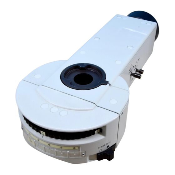
Table of Contents
Advertisement
BX-URA2
BX-RFA
U-LH100HGAPO
U-LH100HG
Power Supply Unit
U-25ND6-2
U-25ND25-2
U-25ND50-2
U-RSL6
U-RSL6EM
BX-RFSS
U-EXBABG
U-EXBAUB
U-EXBAUG
This instruction manual is for the Olympus Reflected Fluorescence System. To ensure the safety,
obtain optimum performance and to familiarize yourself fully with the use of this system, we recom-
mend that you study this manual thoroughly before operating the microscope. Retain this instruc-
tion manual in an easily accessible place near the work desk for future reference.
This publication is printed on 100% recycled paper
INSTRUCTIONS
REFLECTED
FLUORESCENCE
SYSTEM
A X 7 6 2 5
Advertisement
Table of Contents
Troubleshooting

Summary of Contents for Olympus BX-URA2
- Page 1 FLUORESCENCE SYSTEM This instruction manual is for the Olympus Reflected Fluorescence System. To ensure the safety, obtain optimum performance and to familiarize yourself fully with the use of this system, we recom- mend that you study this manual thoroughly before operating the microscope. Retain this instruc- tion manual in an easily accessible place near the work desk for future reference.
-
Page 3: Table Of Contents
CONTENTS Correct assembly and adjustments are critical for the reflected fluorescence system to exhibit its full performance. If you are going to assemble the reflected fluorescence system yourself, please carefully read section 9, “ASSEMBLY” (pages 30 to 35). IMPORTANT — Be sure to read this section for safe use of the equipment. — I. - Page 4 — See this section for the replacement of the light bulb. — 9-1 Assembly Diagram .................................... 30 9-2 Detailed Assembly Procedures ..........................31-35 II. REFLECTED OBSERVATIONS (BX-URA2 Only) CONFIGURATION OF REFLECTED OBSERVATION SYSTEM ASSEMBLY FIELD IRIS AND APERTURE IRIS DIAPHRAGM ADJUSTMENTS...
-
Page 5: Safety Precautions
6. Do not attempt to open or disassemble the power supply unit because it includes high voltage parts inside. 7. Always use the power cord provided by Olympus. If no power cord is provided, please select the proper power cord by referring to the section “PROPER SELECTION OF THE POWER SUPPLY CORD”... - Page 6 Safety Symbols The following symbols are found on the microscope. Study the meaning of the symbols and always use the equipment in the safest possible manner. Symbol Explanation Indicates the presence of high voltage (1 kV or more). Take caution to guard against electric shock.
-
Page 7: Maintenance And Storage
Maintenance and Storage 1. To clean the lenses and other glass components, simply blow dirty away using a commercially available blower and wipe gently using a piece of cleaning paper (or clean gauze). If a lens is stained with fingerprints or oil smudges, wipe it gauze slightly moistened with commercially available absolute alcohol. - Page 8 Fluorescence Illuminator BX-RFA 100 W Mercury Lamp Housing U-LH100HG Note The diagram shows the BX-RFA. Parts marked * are not provided on the BX-URA2. Field iris diaphragm centering screws (Page 12) x2 screws. Collector lens focusing knob (Page 14) Burner centering knobs...
- Page 9 }Up to six fluorescence mirror units can be mounted on the BX-RFA or BX- Fluorescence Mirror Units URA2. U-MWU2, etc., total 24 models # Each filter unit includes a dichroic mirror, barrier filter and excitation filter that have been combined according to the excitation method. It is basically not recommended to open a fluorescence mirror unit.
- Page 10 REFLECTED FLUORESCENCE OBSERVATION PROCEDURE }If you need simultaneous observation of reflected fluorescence observation with the phase contrast observation or trans- mitted light Nomarski Differential Interference Contrast (DIC) observation, please read Chapter 4, “SIMULTANEOUS FLUO- RESCENCE OBSERVATION”. (Page 16) (Controls Used) (Page) Preparation ·...
- Page 11 † ‡ ‹ ‰ ƒ Š ³ ² … } Make a photocopy of the observation procedure pages and post it near your microscope.
-
Page 12: Using The Controls
U-MBF3. }Use according to the excitation wevelength: Olympus has prepared some sets of fluorescence mirror unit combined with appropriate filters which are variable de- pending on wavelengths. The wide-band (W) set is normally used. There may be cases, however, where superwide-band (SW) or Narrow-band (N) sets are recommendable. - Page 13 Dichroic Mirror and Filter Configurations of Fluorescence Mirror Units Excitation Mirror Unit Dichroic Mirror Excitation Filter Barrier Filter Fluorochromes Method · Autofluorescence observation U-MWU2 BP330-385 DM400 BA420 · DAPI: DNA staining U-MNU2 BP360-370 · Hoechest 33258, 33342: Chromosome · Catecholamine DM455 BP400-410 BA455...
-
Page 14: Objectives For Various Observation Modes
Exclusively for Fluorescent Proteins Excitation Mirror Unit Dichroic Mirror Excitation Filter Barrier Filter Fluorochromes Method U-MCFPHQ DM450HQ BP425-445HQ BA460-510HQ For ECFP U-MGFPHQ DM485HQ BP460-480HQ BA495-540HQ For EGFP U-MYFPHQ DM505HQ BP490-500HQ BA515-560HQ For EYFP U-MRFPHQ DM565HQ BP535-555HQ BA570-625HQ For RFP Mirror Unit Name Meaning U - M C F P H Q High Quality CFP/GFP/YFP/RFP... -
Page 15: Turning The Power Supply Unit On
UIS Series Reflected light fluorescence Phase contrast Transmitted Objective difference light DIC U, V, BV B, IB, G, IY UPlanApo ¦ ¦ – – ¦ ¦ ¦** ¦ 10X O ¦ ¦ – ¦ 10X W ¦ ¦ – – ¦... -
Page 16: Centering The Field Iris Diaphragm
Centering the Field Iris Diaphragm (Fig. 1) ² 1. Close the light path by sliding the shutter knob @ to position marked {. ³ 2. Engage the B or IB mirror unit in the light path by rotating the turret. (If these mirror units are not available, engage another fluorescence mir- ror unit in the light path.) 3. -
Page 17: Centering The Aperture Iris Diaphragm
Centering the Aperture Iris Diaphragm (Fig. 2) ³ 1. Close the light path by sliding the shutter knob @ to position marked {. 2. Engage the B or IB mirror unit in the light path by rotating the turret. (If these mirror units are not available, engage another fluorescence mir- ²... -
Page 18: Centering The Mercury Burner
Centering the Mercury Burner ² ³ }Set the main switch to “ I ” (ON) and wait for 5 to 10 minutes until the arc ƒ stabilizes before proceeding to the mercury burner centering. 1. Close the light path by sliding the shutter knob @ to position marked {. 2. -
Page 19: Mounting The Nd Filters
Precise Centering of the Mirror Arc Image }The mirror arc image position has been adjusted and fixed at the factory. … Perform the centering of the mirror arc image after completing the center- ing of the mercury burner and only when you want to make your adjust- ments very strict and precise. -
Page 20: Simultaneous Fluorescence Observations
SIMULTANEOUS FLUORESCENCE OBSERVATIONS }By properly combining equipment, this system can be used in transmitted light brightfield observation, transmitted phase contrast observation and transmitted light DIC observation in addition to the reflected fluorescence observation. With specimens that fade rapidly, fading can be minimized by initially using transmitted light phase contrast or transmitted light DIC observation for positioning. -
Page 21: Troubleshooting Guide
Under certain conditions, performance of the unit may be adversely affected by factors other than defects. If problems occur, please review the following list and take remedial action as needed. If you cannot solve the problem after checking the entire list, please contact your local Olympus representative for assistance. Problem... -
Page 22: Spectral Characteristics Of Filters
SPECTRAL CHARACTERISTICS OF FILTERS 1. U-excitation (Wide band) 4. BV-excitation (Wide band) U-MWBV2 U-MWU2 Wavelength (nm) Wavelength (nm) 2. U-excitation (Narrow band) 5. BV-excitation (Narrow band) U-MNU2 U-MNBV2 Wavelength (nm) Wavelength (nm) 3. V-excitation (Narrow band) 6. B-excitation (Wide band) U-MNV2 U-MWB2 Wavelength (nm) - Page 23 7. B-excitation (Narrow band) 10. IB-excitation (Narrow band) U-MNB2 U-MNIB3 BP470-495 DM505 BA510IF Wavelength (nm) Wavelength (nm) 8. B-excitation (Superwide band) 11. G-excitation (Wide band) U-MSWB2 U-MWG2 Wavelength (nm) Wavelength (nm) 9. IB-excitation (Wide band) 12. G-excitation (Narrow band) U-MWIB3 U-MNG2 BP460-495 DM505...
- Page 24 13. G-excitation (Superwide band) 16. U-excitation, color separation (Narrow band) U-MSWG2 U-MNUA2 Wavelength (nm) Wavelength (nm) 14. IG-excitation (Wide band) 17. IB-excitation, color separation (Wide band) U-MWIG3 U-MWIBA3 BP530-550 BP460-495 DM570 DM505 BA510-550 BA575IF Wavelength (nm) Wavelength (nm) 15. IY-excitation (Wide band) 18.
- Page 25 19. G-excitation for color separation (Wide band) 22. For green fluorescent protein (GFP) U-MWIGA3 U-MGFPHQ BP530-550 DM485HQ BP460-480HQ DM570 BA495-540HQ BA575-625 Wavelength (nm) Wavelength (nm) 20. G-excitation for color separation (Narrow band) 23. For yellow fluorescent protein (YFP) U-MNIGA3 U-MYFPHQ BP540-550 DM505HQ BP490-500HQ...
- Page 26 Typical Example of Emission Spectrum of Ultra-High-Vacuum Mercury Burner Spectrum For fluorochrome emission, a light beam having a specific wavelength is selected from a wide spectrum of wavelengths. The five major peaks of luminance are at wavelengths of 365/366, 404.7, 435, 546.1 and 577.0/579.1 nm. In addition, light beams having wave- lengths of 334.2 and 490 nm (at rather low luminance) are also applicable to fluoro- chrome emission.
-
Page 27: Specifications
Item Specification Vertical illuminators Reflected Illuminator Fluorescence Illuminator BX-URA2 BX-RFA · UIS2/UIS (Universal Infinity System) optical system (featuring infinity correction) · Magnification: 1X (Superwide field: NA 26.5) · Observation switching: Mirror unit turret carrying max. 6 mirror units. · Aperture iris diaphragm and field iris diaphragm (Both centerable) Detachable with the BX- RFA. -
Page 28: Optional Modules
6-Position Filter Slider U-RSL6 }This filter slider is for use with the BX-URA2 or BX-RFA illuminator and accommodates a total of six excitation and ND filters. It is designed to prevent centering deviation between the optical axes of the excitation filters when multiple excitation mirror units are used and switched. - Page 29 Using the Joint Plates The joint plates @ can be attached and locked between the slider knob and slider as shown in the figure. The joint plates should be attached on both ends of the filter slider. By locking with the joint plates, you can switch the barrier and excitation filters together as a set.
-
Page 30: 6-Position Barrier Filter Slider U-Rsl6Em
6-Position Barrier Filter Slider U-RSL6EM }This filter slider is for use with the BX-URA2 or BX-RFA illuminator and accommodates a total of six barrier filters. Slider knob Joint plate Light shield plate (x 4) Inscription sticker position Diameter 8 mm, depth 1 mm... -
Page 31: Rectangle Field Stop Bx-Rfss (For Exclusive Use With The Bx-Rfa)
Rectangle Field Stop BX-RFSS (for exclusive use with the BX-RFA) }When fluorescence images are recorded with the TV camera for observation or image processing, this unit projects a rectangular iris diaphragm image with size variable according to the captured image size. This helps prevent color fading of specimen due to other reasons than image capturing. -
Page 32: Exciter Balancers U-Exbabg/Exbaub/Exbaug (For Exclusive Use With The Bx-Rfa)
Observing a Double Stained Specimen 1. Set up normal reflected fluorescence observation. 2. Mount the fluorescence mirror units for double staining and engage them in the light path. }Olympus standard products # Due to its own characteristics, the G-exci- Exciter Balancer... - Page 33 3. Push in the adjustment lever of the balancer slider to be used to engage the filter in the light path. }The angle of each adjustment lever can be adjusted in the range shown below, only when the lever is pushed in. 0˚...
-
Page 34: Assembly
) provided with the illuminator is used only for clamping the screws inside the illuminator. (To retain the performance, have your Olympus representative conduct this work.) · Parts marked with # can be attached only to the BX-URA2 universal illuminator. NOTES ·... -
Page 35: Detailed Assembly Procedures
9-2 Detailed Assembly Procedures Attaching the Fluorescence Mirror Units (Figs. 10 & 11) 1. Using the Allen screwdriver, loosen the clamping screw @ at the right side of the vertical illuminator. 2. Pull out the turret and place it upside down. }Dummy mirror units ²... - Page 36 Making an Optional Fluorescence Mirror Unit }You can also fabricate optional fluorescence mirror units by fitting a commercially available barrier filter, excitation filter or dichroic mirror in the U-MF2 mirror unit frame. Dimensions of Optical Parts · Barrier filter Diameter 25 -0.1/-0.2 mm, max. thickness 6 mm. ·...
- Page 37 Attaching the Mercury Burner (Figs. 12 - 15) 1. Loosen the socket clamping screw @ using the Allen screwdriver. 2. Hold the upper section of lamp housing and pull it upward to remove the socket section. ² # To prevent malfunctions, do not hold the lamp housing by the cen- tering knobs ².
- Page 38 Resetting the Burner Hour Counter 1. Press the center section @ of the reset button ² on the power supply unit’s front panel to reset the hour counter to 000.0. }The hour counter shows elapsed time in hours. The service life of a burner is 300 hours.
- Page 39 !Make sure that the main switch is set to “ ” (OFF) before connect- ing the power cord. !Always use the power cord provided by Olympus. If no power cord ² is provided, please select the proper power cord by referring to ³...
-
Page 40: Reflected Observations (Bx-Ura2 Only)
CONFIGURATION OF REFLECTED OBSERVATION SYSTEM The BX-URA2 universal illuminator can be used in a variety of brightfield observations, darkfield observation, DIC observation and simplified polarized observation under reflected lighting when it is used in combination with a UIS2/UIS objective for metallic specimens, the U-MBF3 brightfield mirror unit, U-MDF3 darkfield mirror unit, etc. -
Page 41: Assembly
ASSEMBLY }This chapter pertains only to the assembly of items which cannot be assembled in the same way as the fluorescence modules. Attaching the U-RCV Conversion Lens (Fig. 18) }Be sure to use this conversion lens when the U-MDF3 mirror unit for darkfield observation is used. - Page 42 4. Fit the Allen screwdrivers provided with the microscope frame into the Field iris image two field iris diaphragm centering screws ³ and adjust them so that the field iris image of the diaphragm is centered on the field of view. 5.
-
Page 43: Observations
OBSERVATIONS 4-1 Reflected Light Brightfield/Darkfield Observations Selecting the Light Path for Observation (Fig. 22) Rotate the turret @ to set the mirror unit matching the required observa- tion method in the light path. Inscription Mirror Unit Field Iris Aperture Iris Reflected light brightfield U-MBF3 Adjust as required. -
Page 44: Reflected Light Nomarski Differential Interference Contrast (Dic) Observation
Reflected Light Nomarski Differential Interference Contrast (DIC) Observation # The performance of polarizer may deteriorate when it has been exposed to light for a long period (about continuous 2000 hours). If this happens, replace the polarizer. # When using the high-intensity light source, be sure to use the U-25L42 filter for prevention of the polarizer burn. }When performing sensitive color observation using the U-DICRH DIC slider, combine the U-POTP3 polarizer. -
Page 45: Observation Procedure
3. With the U-DICRH or U-DICRHC slider that does not have the slide lever, the applicable objectives are as follows. DIC Slider Applicable Objectives UIS2 U-DICRH MPLFLN/MPLFLN-BD series UMPlanFl/UMPlanFl-BD series MPlanFl-BD series MPlanApo20X, 100X UIS2 U-DICRHC LMPLFLN/LMPLFLN-BD series LMPlanFl/LMPlanFl-BD series LMPlanApo/LMPlanApo-BD series Observation Procedure 1. -
Page 46: Reflected Light Simple Polarized Light Observation
Switching Between Brightfield and Darkfield Observation 1. Loosen the DIC clamping screw @ at the front of the revolving nose- piece, and gently pull the DIC prism ² outward until a click is heard. Tighten the claming screw again. 2. Rotate the turret to disengage the U-MDIC3 DIC mirror unit from the light path. -
Page 47: Optical Characteristics
(as shown in the diagram on the right). UIS marking NA (Numerical Aperture) NOTE Brightfield/darkfield Refer to the latest catalogue or consult Olympus for the updated infor- application mation on the eyepieces and objectives that can be combined with this unit. Cover glass thickness FN (Field Number) —: May be used with our without... - Page 48 Significance of Objective Name (Examples) (Plan) None : Brightfield : Brightfield/darkfield BDP : Brightfield/darkfield or polarized : IR light Figure : Magnification None : UIS : UIS 2 None : Achromat, or aberration correction with 2 wavelengths (red and bleu). : Semi-Apochromat, or color aberration correction with visual wave- lengths (bluish purple to red).
-
Page 49: Troubleshooting Guide
TROUBLESHOOTING GUIDE Reflected Light Observation Modes Problem Cause Remedy Page a) Bulb operates, but field of view re- Reflected light lamp is not on. Turn lamp on. mains dark. Aperture and field iris diaphragms are not Enlarge them to proper sizes. opened wide enough. -
Page 50: Proper Selection Of The Power Supply Cord
If no power supply cord is provided, please select the proper power supply cord for the equipment by referring to “ Specifications ” and “ Certified Cord ” below: CAUTION: In case you use a non-approved power supply cord for Olympus products, Olympus can no longer warrant the electrical safety of the equipment. - Page 51 Table 2 HAR Flexible Cord APPROVAL ORGANIZATIONS AND CORDAGE HARMONIZATION MARKING METHODS Alternative Marking Utilizing Printed or Embossed Harmoniza- Black-Red-Yellow Thread (Length tion Marking (May be located on Approval Organization of color section in mm) jacket or insulation of internal wir- ing) Black Yellow...
- Page 52 Shinjuku Monolith, 3-1, Nishi Shinjuku 2-chome, Shinjuku-ku, Tokyo, Japan Postfach 10 49 08, 20034, Hamburg, Germany 3500 Corporate Parkway, P.O. Box 610, Center Valley, PA 18034-0610, U.S.A. Vision House, 19 Colonial Way, Watford, Hertfordshire, WD24 4JL, UK One Corporate Drive, Orangeburg, NY 10962, U.S.A. 491B River Valley Road, #12-01/04 Valley Point Office Tower, Singapore 248373 31 Gilby Road, Mount Waverley, VIC., 3149, Australia 5301 Blue Lagoon Drive, Suite 290 Miami, FL 33126, U.S.A.


