Sign In
Upload
Manuals
Brands
DRAMINSKI Manuals
Medical Equipment
4Vet Slim
DRAMINSKI 4Vet Slim Manuals
Manuals and User Guides for DRAMINSKI 4Vet Slim. We have
1
DRAMINSKI 4Vet Slim manual available for free PDF download: User Manual
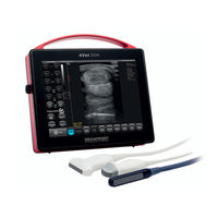
DRAMINSKI 4Vet Slim User Manual (60 pages)
Diagnostic ultrasound scanner with a touch panel
Brand:
DRAMINSKI
| Category:
Medical Equipment
| Size: 3 MB
Table of Contents
Table of Contents
4
1 Introduction
8
Information about User Manual
8
Warnings and Comments Used in this User Manual
8
Brief Information on Ultrasound Examination
9
Basic Information about the Ultrasound Scanner
9
2 User's Safety
10
3 The List of DRAMIŃSKI 4Vet Slim Elements and Equipment
11
4 Construction of DRAMIŃSKI 4Vet Slim Ultrasound Scanner
11
The Casing
11
Ultrasound Probe
13
DRAMIŃSKI Battery Pack
13
Battery Charger
14
AC Adapter
14
5 Technical Data
15
6 Preliminary Starting Procedure
16
General Principles of Using the Touch Panel
17
Connecting Battery Pack
17
Connecting the Probe
18
Starting the Device Using the Mains Supply
19
Starting the Device Using Battery Power Supply
19
Finishing Work
19
Transportation of the Device
20
7 Preparation to Perform Ultrasound Examination
20
8 Description of the User Panel and Available Functions
21
Basic Panel (B, B+B, B+M Modes)
21
Active Fields
23
Panel in Color Doppler Mode
24
Power Doppler Mode Panel
25
Pulse Wave Doppler Mode Panel
26
9 Imaging Modes
27
Choosing Imaging Mode
27
B, B+B and 4B Modes
27
Switching Windows between B+B and 4B Modes
27
M and B+M
28
Doppler Modes: Color Doppler, Power Doppler and Pulse Wave Doppler
28
Switching between Color Doppler, Power Doppler and Pulse Wave Doppler Modes
28
10 Description of the Ultrasound Scanner Functions
28
Optimizing the Image Parameters
28
Adjustment of Signal Gain [Gain 1] and [Gain 2]
28
Selection of the Probe's Frequency
29
Change of the Penetration Depth
29
Focus-Setting (Focusing the Beam)
29
Zoom
29
Gamma Level Correction
30
Invert Color
30
Freeze
30
Cine Loop - Continuous and Frame-By-Frame Replay
30
Measurements
31
Length
31
Grid
32
Stenosis
32
Volume
32
Area
33
The Area of Ellipse
33
Gynecological and Obstetric Measurements
34
Horse DC] - Horse Head Circumference
34
Horse DO] - Diameter of Eyeball
34
Horse DSG] - Horse Gestational Sac Diameter
34
Sheep LCC] - Sheep Crown - Rump Length
34
Lama BPD] - Lama Biparietal Diameter
34
Sows LCC] - Pig Crown - Rump Length
34
Cow BPD] - Cow Biparietal Diameter
34
Cow LCC] - Cow Crown - Rump Length
34
DOG GS Small] - Gestational Sac Diameter - Small Dog
34
DOG BPD Medium] - Biparietal Diameter - Medium Dog
35
Cat >30 HD] - Head Diameter - Cat
35
Cat >30 BD] - Body Diameter - Cat
35
Cat <30 CRL] - Crown - Rump Length - Cat
35
Cat <30 GSD] - Gestational Sac Diameter - Cat
35
Edition of the Measurements
36
Clear]
36
Optimization of B+M and M Modes
37
Graph Drawing Speed Selection in M Mode
37
Setting the Cutting Line in B+M / M Modes
37
Switching between M and B+M Modes
37
Imaging with the Function of Color Doppler
37
Starting Color Doppler Imaging
37
Switching between the Doppler Modes: Color, Power, Pulse Wave; Doppler Mode off
37
Setting Doppler Frame
38
Repositioning of Doppler Frame
38
Resizing of Doppler Frame
38
Tilt Adjustment of Doppler Frame for Linear Probe
39
Setting Parameters for Doppler Mode
39
PRF] - Pulse Repetition Frequency
39
Mhz] - Frequency
39
Gain] - Color Adjustment
39
Averaging]
39
Persistence]
40
Angle] - Setting Tilt Angle of Doppler Frame
40
Color Invert]
40
Color Threshold]
40
Color Range]
41
Power Doppler (Color Coding Power Flows Doppler)
41
Setting Power Doppler
41
Postprocessing
41
Filtered / Not Filtered Averaging
41
Pulse Wave Doppler (Spectral Pulse Doppler)
41
Setting Parameters in Pulse Wave Doppler Mode
41
Depth -/+ ] (Sampling Gate Depth)
42
Insonation Angle]
42
Width -/+ ] (Sampling Gate Width)
42
Spectrum Registration of Blood Flow
42
Modification of Spectral Graph
42
Measurement in Pulse Wave Doppler Mode
42
HR] - Heart Rate
43
AT] - Acceleration Time
43
Ri Pi Psv Edv]
43
Doppler Point]
43
Saving and Loading the Images and Cine Loop to the Screen
43
Saving the Image
43
Saving the Cine Loop
44
Loading the Images
44
Loading Cine Loop
45
Filtering Stored Data
46
Edition of Stored Images and Cine Loops
46
Data Export to External Storage Medium
46
Data Export in DICOM Format
46
Panoramic Image (Option)
46
Function "Patient
47
Retrieve Data from DICOM Worklist
47
Function "Needle
48
Full Screen Function
48
Image Printing
48
Printing During Examination
48
Printing of Stored Images
49
Printing Multiple Images on One Sheet
49
Presets
49
Presets Activation
49
Presets Saving
49
11 Advanced Settings
50
View
50
Option „One Button" for Save and Load Files
51
Clinic
52
Info
52
Software Update
52
On-Line Update
52
Update Using USB Flash Drive
52
Choosing Printer
53
Choosing the File Name Creation Style of the Exported Files
53
Date and Time Settings
53
Display
53
Measurements Settings
53
DICOM Settings
53
DICOM Printer Configuration
53
MWL] - Modality Worklist Configuration
54
Configuration of Data Export to the DICOM Server
54
12 Charging and Exploitation of the Batteries
54
Charging DRAMIŃSKI Battery Pack
55
13 Maintenance of the Device
55
14 Troubleshooting
57
15 Symbols and Marks Used on the Labels
58
16 Warranty
59
Advertisement
Advertisement
Related Products
Draminski 4Q Mast
Draminski 4x4Q Mast
Draminski BLUE
Draminski DermaMed
Draminski FAST
DRAMINSKI Categories
Measuring Instruments
Security Sensors
Scanner
Medical Equipment
Diagnostic Equipment
More DRAMINSKI Manuals
Login
Sign In
OR
Sign in with Facebook
Sign in with Google
Upload manual
Upload from disk
Upload from URL