
Related Manuals for Norland Elite 437A150

Summary of Contents for Norland Elite 437A150
- Page 1 DXA Bone Densitometer Operator’s Guide Elite Model 437A150 Operator's Guide 437D140 Rev. I...
- Page 2 All Norland Products are covered by U.S. and other patents and/or patents pending. All product names are trademarks of Norland. All trademarks and the rights of the trademarks owned by the companies referred to in this Operator’s Guide and other written materials provided to Licensee with the Bone Assessment Product are acknowledged.
- Page 3 Software. Licensee acknowledges that Norland retains proprietary rights in the Soft- ware. In the event of any misappropriation or misuse, in addition to its right to damages Norland shall have the right to obtain injunctive relief. Licensee agrees not to decompile, disassemble or reverse engineer the Software.
- Page 4 New York County and to any other court in any jurisdiction in which an action is brought against a party to this agreement by a third party asserting a claim against which Norland is entitled under this agree- ment to be indemnified.
-
Page 5: Table Of Contents
Contents Introduction Indications for Use Contraindications X-Ray Device Registration Customer Service Contact Information About This Manual Symbols List ON/OFF Power Switch Scanner Arm Touch Pad Symbols General Information Radiation Safety Precautions Stay Out of the Beam Avoid Scatter Radiation State Requirements Control General Access Radiation Safety Features Exposure Control... - Page 6 Contents Editing the Patient Letter or Referral Letter 2-16 Sample Scan Reports 2-17 The Bone Exam Report 2-17 1: A Sample “Bone Exam Report” 2-18 2: A Sample “Bone Exam Report - 1 Page” 2-20 3: A Sample “Combined Report” 2-21 4: A Sample “Patient Letter”...
- Page 7 Patient Scanning Procedure Overview Guidelines for Attaining Precision and Accuracy Powering Up the System Logging into the Norland Software Daily Calibration Procedure 1. Check the Standard and QC Phantom for Damage 2. Position the Standard and the Phantom on the Scanner 3.
- Page 8 Contents Marking the Scan Region 5-10 Starting the Measure Scan 5-13 Analyzing the Scan 5-15 Viewing the Scan Results Tab 5-18 Generate and Print a Report 5-20 A Sample Bone Exam Report 5-22 Soft Tissue Composition Introduction Tissue Composition Standards Scan Specifications Whole Body Scans Soft Tissue Composition - Whole Body Scans...
- Page 9 Contents Checklist Preparing the Patient for Scanning Update (or Create) the Patient’s Record Setting the Scan Parameters Positioning the Patient 8-10 Marking the Scan Region 8-16 Starting the Scout Scan 8-18 Starting the Measure Scan 8-21 Analyzing the Scan 8-23 Viewing the Scan Results Tab 8-26 Definitions of Scan Results...
- Page 10 viii Contents Starting the Measure Scan 10-19 Analyzing the Scan 10-21 Viewing the Scan Results Tab 10-24 Generate and Print a Report 10-26 A Sample Bone Exam Report 10-28 Additional Techniques Adding Audio to a Patient Record 11-2 Using the Voice Note Option 11-3 How to Record a Voice Note 11-3...
- Page 11 Contents Comparison Image 11-54 Image Composition and Image Mode 11-56 Analyzing Saved Scan Data 11-59 Reanalyzing Scan Data 11-61 Changing Height or Weight on the Report 11-62 AP Spine Techniques 11-63 Changing the Scan Parameters Prior to Scanning 11-63 Unsatisfactory Measure Scan (AP Spine) 11-64 Auto Centering Mode 11-65...
- Page 12 Contents Modality Value 13-8 Quick Reference - DICOM Interface 13-9 Patient Worklist 13-9 Exporting and Pushing DICOM Reports 13-9 Patient Worklist 13-10 Query / Cancel 13-11 1. No Results 13-11 2. Error in Query 13-11 3. Query Results Area 13-11 Data Patient Conflict 13-13 Exporting DICOM Images...
- Page 13 Calibration 16-6 Printer 16-7 Reference/Trend Chart Display 16-8 Height and Weight on Reports 16-9 FolderMAIL 16-9 Technical Reference Norland Bone Densitometer System Specifications 17-2 DXA System Description 17-2 Electrical Requirements 17-2 Environmental 17-2 X-Ray Detector Assembly 17-2 Scanner Arm Drive...
- Page 14 Norland Fracture Risk 18-6 Excerpt from Calcified Tissue International 18-7 Excerpt from the Journal of Bone and Mineral Research 18-8 Norland Reference Data Set Values 18-10 NHANES III Reference Data Set Values 18-15 Index Quick Reference - AP Spine Scan...
- Page 15 ISO 13485 Certified: Norland’s Quality Management System is certified to ISO 13485:2003. Norland is dedicated to providing a quality product, on time delivery, and exceptional customer service. Caution: State law requires all scans to be prescribed by a physician.
-
Page 16: Introduction
Mass, Fat Mass, % Total Fat, and the % Soft Tissue Fat for the Whole Body, Research and Small Subject scans. It also provides Siri and Brozek (underwater weighing) % Fat equivalent values. The bone density measurements from the Norland Bone Densitometer can be an aid to physicians in determ- ining a patient’s risk of fractures. -
Page 17: Customer Service Contact Information
All requests for replacement parts, service or product related information should be directed to one of the fol- lowing worldwide locations. To help the Norland representatives provide prompt and efficient service, please have the serial number of the instrument available. See the figure on on page 17-17 (of the Technical Reference chapter) for the label location. -
Page 18: About This Manual
Chapter 9, Research & Small Subject Scan: as options, these two types of scans are very similar, and are therefore combined into one chapter. The Norland software quantifies bone mineral in any user-defined region of a patient or subject anywhere within the scanner’s active scanning area. The subject of a Research or Small Subject scan could be human, animal, or an inanimate object. - Page 19 Labels are discussed in detail. Chapter 18, Reference Data Sets: contains information about the Norland Reference Data Sets, including the data collection process and criteria. A listing of the values of each Reference Data Set is also included in this chapter.
-
Page 20: Symbols List
Introduction Symbols List Indicates information, general caution or possible safety hazard. Indicates helpful notes or additional procedure steps. Indicates radiation from laser positioning aid. ON/OFF Power Switch Turn the scanner ON and OFF by depressing the power switch located on the lower inside right panel. Power Switch location Figure 1-1: Operator's Guide... -
Page 21: Scanner Arm Touch Pad Symbols
Introduction Scanner Arm Touch Pad Symbols Figure 1-2: Scanner Touch Pad The Radiation exposure and arm movement can be terminated, and the system power turned off imme- diately by the operator at any time by pressing the EMERGENCY STOP button located directly below the Scanner Arm Touch Pad. - Page 22 This page intentionally left blank. Operator's Guide 437D140 Rev. I...
-
Page 23: General Information
The Norland DXA Bone Densitometer uses a technique known as Dual Energy X-Ray Absorptiometry (DXA) to perform non-invasive estimates of bone mineral density (BMD) in specific regions of the body. The bone density estimates from the Norland DXA system can be used to aid the physician in diagnosing and managing osteo- porosis, as well as an aid in assessing risk of fracture. -
Page 24: Radiation Safety Precautions
General Information Radiation Safety Precautions Radiation safety is an important consideration whenever working with x-ray devices. The Norland system emits very little radiation to the patient or scatter radiation to the operator, however certain precautions should be followed to ensure a safe environment for operators and patients. -
Page 25: Radiation Safety Features
At the typical distance of 3 feet from the beam, the radiation level is <0.1 mRem/hour. Norland recommends that operators position them- selves at least three feet from the beam during the scan or as indicated by local state x-ray regulations. -
Page 26: Dual Energy X-Ray Absorptiometry (Dxa)
Norland uses samarium as the filter material because it produces energy peaks at 46.8keV and 80keV, which have proven to be most effective at differentiating between soft tissue and bone tis- sue. -
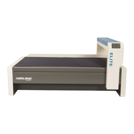
Page 27: System Components
General Information System Components The Norland Bone Densitometer consists of a Scanner Unit and a Control/Analysis Computer with a color printer. Scanner Unit The Scanner Unit is a specially constructed table that accommodates the patient in a supine position. The tab- letop is cushioned and upholstered with a highly durable antimicrobial fabric that can be removed for cleaning. -
Page 28: Laser Positioning Aid
Do not allow shiny objects to reflect laser light into your eyes. Computer/Controller The Norland Bone Densitometer is controlled by a desktop PC. The computer is designed and configured to operate the Norland Bone Densitometer system and should not be used for any other purpose. The computer is certified as an X-Ray Controller at Norland's manufacturing facility, as required by the FDA. -
Page 29: Patient Positioning Aids
General Information Patient Positioning Aids Patient positioning aids are provided for special positioning of the patient during spine, hip, and lateral pro- cedures. Table 2-1: Patient Positioning Aids Positioning Aid Part Number Description Hip Sling 433A134 Used to orient the patient's legs in a position best suited for accurate and precise hip scans. -
Page 30: Quality Assurance Program
Sample pop-up: Figure 2-3: QA Alert pop-up It is not necessary to calibrate the system on a day that the Norland Bone Densitometer will not be used to scan patients. Patient Comparison Patient scan results are compared to reference populations or to previous scans for comparisons for diagnosis. -
Page 31: Trend Comparison
Precision is the degree to which the same value is obtained when a measurement is repeated. In vivo precision in the Norland Bone Densitometer is the degree to which the device presents the same bone mineral value when a measurement is repeated at the same anatomical site on the same subject. -
Page 32: Accuracy
The accuracy spe- cifications for the Norland Bone Densitometer are stated in terms of accuracy of the mean. Several manufacturers have adopted phantoms fabricated of calcium hydroxyapatite and epoxy. The cal- ibration of each Norland instrument is based on hydroxyapatite phantoms constructed to specifications described by White (White, D. - Page 33 High Risk (Red) Figure 2-4: Fracture Risk Reference Set Chart (sample) Norland incorporates the WHO (World Health Organization) criteria in plotting a patient's fracture risk assess- ment (see table below). Table 2-2: World Health Organization Risk Factor Criteria Low Risk...
-
Page 34: Scan Reports
2-12 General Information Scan Reports Five different types of Reports can be generated for each scan: the Bone Exam Report , the Bone Exam Report - 1 Page , the Combined Report , the Patient Letter , and the Referral Letter . In addition, the Ten Year Fracture Risk Report can be generated for Hip Scans (if enabled), and the Body Composition Report can be generated for Whole Body Scans (if enabled). - Page 35 General Information 2-13 5. The Report is immediately generated and opens up in the window, as shown. At this point, the operator can type in their comments and recommendations, Lock it, Save it, Print it, Export it to a PDF file (or a DICOM file) and Close it.
-
Page 36: The "Reports" Tab
2-14 General Information The “Reports” Tab As mentioned previously, once a Report is generated and saved, it can be viewed from the “Reports” tab. It can be modified, viewed and re-printed at any time. 1. From the Stamp view, click the Reports tab to see the Report list (shown here). 2. - Page 37 General Information 2-15 To modify a locked Report, click on Tools > Report > Make Addendum. Make any edits to the Comments field as necessary. Click , then Operator's Guide 437D140 Rev. I...
- Page 38 2-16 General Information Editing the Patient Letter or Referral Letter The information from Bone History and Osteoporosis Medication fields in the Demographics screen will be lis- ted in the Referral Letter. Scan Quality Issues and Recommendations can be added to either report using a checklist. To edit the Scan Quality Issues checklist click on Tools >...
-
Page 39: Sample Scan Reports
General Information 2-17 Sample Scan Reports The Bone Exam Report The results of the Analysis function include image displays, graphical displays and numeric information and are typically printed as a two page report. This report is referred to as the “Bone Exam Report”. An example of a Bone Exam Report is included in this section. -
Page 40: 1: A Sample "Bone Exam Report
2-18 General Information 1: A Sample “Bone Exam Report” A sample 2-page Bone Exam Report is included here for reference. Figure 2-5: Page 1 of the Bone Exam Report Operator's Guide 437D140 Rev. I... - Page 41 General Information 2-19 Page 2 of the Bone Exam Report Figure 2-6: Operator's Guide 437D140 Rev. I...
-
Page 42: 2: A Sample "Bone Exam Report - 1 Page
2-20 General Information 2: A Sample “Bone Exam Report - 1 Page” The information from the 2-page Bone Exam Report is condensed down to form this one page report, with basic patient information and reference graphs for all regions of interest, as shown next: Figure 2-7: The Bone Exam Report - 1 Page Operator's Guide... -
Page 43: 3: A Sample "Combined Report
General Information 2-21 3: A Sample “Combined Report” The physician has the option to combine the results of two different bone scans of a single visit into a one page Combined Report, as shown next: Figure 2-8: The Combined Report Operator's Guide 437D140 Rev. -
Page 44: 4: A Sample "Patient Letter
2-22 General Information 4: A Sample “Patient Letter” The Patient Letter is a 2 or 3 page Report that is sent to a patient from the primary physician. Patient Letters can be generated for a single scan or a combination of two different scans from the same visit. “Scan Quality Issues”... - Page 45 General Information 2-23 Page 2 of the Patient Letter: Figure 2-10: Operator's Guide 437D140 Rev. I...
-
Page 46: 5: A Sample "Referral Letter
2-24 General Information 5: A Sample “Referral Letter” The Referral Letter is a 2 or 3 page Report sent to the referring physician from the facility that did the scan. Referral Letters can be generated for a single scan or a combination of two different scans from the same visit. “Scan Quality Issues”... - Page 47 General Information 2-25 Page 2 of the Referral Letter Figure 2-12: Operator's Guide 437D140 Rev. I...
-
Page 48: 6: A Sample "Ten Year Fracture Risk Report
2-26 General Information 6: A Sample “Ten Year Fracture Risk Report” A Ten Year Fracture Risk Report assess the patient's probability of a future fracture. This report can only be generated for hip scans and is only available when the Ten Year Fracture Risk option is enabled. Figure 2-13: Page 1 of the Ten Year Fracture Risk Report Operator's Guide... - Page 49 General Information 2-27 Page 2 of the Ten Year Fracture Risk Report Figure 2-14: Operator's Guide 437D140 Rev. I...
-
Page 50: 7: A Sample "Body Composition Report
2-28 General Information 7: A Sample “Body Composition Report” A Body Composition Report assesses the soft tissue and bone of a Whole Body scan to produce Body Fat Percentage results. This report can only be generated for Whole Body scans and is only available when the Body Composition Reports option is enabled. - Page 51 General Information 2-29 Page 2 of the Body Composition Report with trended results Figure 2-16: Operator's Guide 437D140 Rev. I...
-
Page 52: Quality Assurance Reports
2-30 General Information Quality Assurance Reports Following are sample Quality Assurance Reports. As described later in this manual, daily system calibrations should be performed daily prior to patient scanning to ensure quality bone density estimates. During the procedure, the QA Calibration Standard is measured first. Next, the QC Phantom is scanned. Your system will have either a clear or a black phantom, depending on its model or options. -
Page 53: Sample Reports: Quality Assurance Report - Bmd
General Information 2-31 Sample Reports: Quality Assurance Report - BMD QC Results - BMD Figure 2-17: Operator's Guide 437D140 Rev. I... -
Page 54: Sample Reports: Quality Assurance Report - Fat
2-32 General Information Sample Reports: Quality Assurance Report - Fat QC Results - Fat (an option) Figure 2-18: Operator's Guide 437D140 Rev. I... -
Page 55: Sample Reports: Quality Assurance Report - Lean
General Information 2-33 Sample Reports: Quality Assurance Report - Lean QC Results - Lean (an option) Figure 2-19: Operator's Guide 437D140 Rev. I... - Page 56 2-34 This page intentionally left blank. Operator's Guide 437D140 Rev. I...
-
Page 57: Installation And Setup
Installation and Setup The first part of this chapter includes the installation instructions for the Norland Illu- minatus DXA software. The installation of the microphone and speakers is also dis- cussed. Uninstalling the software concludes the installation section. Instructions for logging on to the system follows next. -
Page 58: Software Installation Instructions
Installation and Setup Software Installation Instructions Installing the IlluminatusDXA Software This procedure assumes that the computer has been set up and is running. If you will be connecting to a net- work, you will need to know your SMTP domain name before beginning the installation. You can get it from your IT person. - Page 59 Installation and Setup 6. Norland recommends that you keep the default Destination Folder. (If you do not, certain files will not be cre- ated correctly and the software will not be able to find them.) Click 7. The application and any selected options will be installed.
- Page 60 Installation and Setup 8. The Sentinel HASP drivers will be automatically installed on the first installation. Click OK when completed. 9. If the Acrobat Reader option was selected, follow the installation prompts to install Acrobat Reader. 10. After all features have been installed, click to complete the installation.
-
Page 61: Configuring Backup For Windows 10
IlluminatusDXA software and data on a daily schedule. To configure the EaseUS backup program on a computer not provided by Norland, follow the steps below. (The EaseUS program may be used on any system running Windows XP or newer.) 1. -
Page 62: Configuring Backup For Windows 7
Configuring Backup for Windows 7 Computers provided by Norland with Windows 7 are configured to automatically perform a backup of the Illu- minatusDXA software and data on a weekly schedule. To configure the backup on a Windows 7 computer not provided by Norland, follow the steps below. - Page 63 Installation and Setup 3. Click on Sounds and Audio Devices. 4. Click the Audio tab. Then click the Sound Recording button. 5. Click the Microphone “Select” check-box then adjust the slide bar to the middle setting. 6. Click the to close the dialog box. 7.
-
Page 64: Uninstalling The Software
Installation and Setup Uninstalling the Software Follow these instructions to uninstall the Illuminatus DXA software, if necessary. 1. Reboot the computer to confirm all programs have been closed. 2. Remove the HASP USB key. 3. Click Start > Control Panel. 4. - Page 65 Selecting Yes will permanently delete all of your patient data and scanner setup files. 9. Wait while the software continues to uninstall the Norland software. 10. Upon completion, the following window opens. Select Yes to restart the computer now or select No to restart later.
- Page 66 3-10 Installation and Setup 11. Click Operator's Guide 437D140 Rev. I...
-
Page 67: Logging Into The Illuminatus Dxa Software
Illuminatus DXA to start the software. OR. . . click on Start > Programs > Norland > Illuminatus DXA. 3. First time login: The following window pops up (after the initial splash screen) when logging in for the first time. -
Page 68: Examining The Software
Norland Bone Densitometry devices are controlled by menu driven software that follows a Windows based design. The main software window is called the Database Navigator window. All Norland program actions are initiated by moving a mouse pointer to the menu bar located along the top of the window or the navigation but- tons along the bottom of the window. -
Page 69: Database Navigator Window
3-13 Database Navigator Window The Norland Bone Densitometer main window is referred to as the Database Navigator window in this manual. It is shown below. All the visits pertaining to one patient are grouped together and sorted under that patient’s name and ID number. -
Page 70: Database Navigator Window Description
3-14 Installation and Setup Database Navigator Window Description The menu bar is located along the top of the window. Click on a menu name to reveal a drop-down list of com- mand names. The commands are: File , Edit , Calibration , Tools , and Help . These commands perform functions in the system software. -
Page 71: Menu Commands Overview
Installation and Setup 3-15 Menu Commands Overview As mentioned earlier, when you click any one of the menu names along the top of the Database Navigator win- dow, a list of commands “drop” down. Some commands are grayed out due to one of the following reasons: The option is not valid for the current operation. -
Page 72: File Command
3-16 Installation and Setup File Command To view the File commands, click on File in the Database Navigator window. Move the pointer down to highlight the command of interest and click on it. The highlighted command will execute. Quick Descriptions: File drop-down list New Patient: displays the Patient Demographics window for inputting patient information. -
Page 73: Edit Command
Installation and Setup 3-17 Edit Command To view the Edit commands, click on the Edit menu in the Database Navigator window. Quick Descriptions: Edit drop-down list User Preferences Main Settings tab: displays dialog box for changing login name and password, e-mail address, and the width and height of the Stamp View thumbnail. - Page 74 3-18 Installation and Setup Users > Main Settings: displays the dialog box for changing login name and password, e-mail address, and the width and height of the Stamp View thumbnail. Users > User’s Groups: contains the Group List. Users > Additional Settings: has Security Privileges settings. Groups tab: contains the Group List.
-
Page 75: User Preferences Command
Installation and Setup 3-19 User Preferences Command Clicking on the User Preferences selection displays tabbed windows that lets the current user change their login and e-mail information. The width and height of the Stamp View thumbnail (that is displayed in the Stamp View window) can be set here, also. -
Page 76: Scanner Preferences Command
3-20 Installation and Setup Scanner Preferences Command The Scanner Preferences selection displays four different tabbed dialog boxes for setting Site information, Scanning parameters, Analysis parameters, and Service Information. Figure 3-2: The Scanner Preferences selection window Most of the scanner parameters will be set during installation prior to applications training. Settings will be dis- cussed with the operator during the training to ensure a complete understanding. -
Page 77: Site Tab Setup
Installation and Setup 3-21 Site Tab Setup The Site tab is accessed by clicking on Edit > Scanner Preferences. Then click the Site tab. The Site tab allows the operator to enter the site and scanner identification information. The available software options are displayed once the Options Authorization Code (OAC) is entered by the installer. -
Page 78: Scanning Tab Setup
3-22 Installation and Setup Scanning Tab Setup The Scanning tab is accessed by clicking on Edit > Scanner Preferences. Then, click the Scanning tab. Here, the operator can set several scan preferences. The scanning preferences available include: AP Spine, Hip, Forearm, Whole Body, Lateral Spine, and Research/Small Subject. They are described in the next few sections. - Page 79 Installation and Setup 3-23 Preferences: AP Spine Scan The following figure shows the default AP Spine Scan parameters. The default settings should be effective for most scanning situations. The default AP Spine Scan Parameters Figure 3-5: Note: WHAT TO DO: To restore the default Scan parameters, click .
- Page 80 3-24 Installation and Setup Preferences: Hip Scan The following figure shows the default Hip Scan settings. The default settings should be effective for most scan- ning situations. Figure 3-6: The default Hip Scan Parameters Note: WHAT TO DO: To restore the default Scan parameters, click .
- Page 81 Installation and Setup 3-25 Preferences: Forearm Scan The following figure shows the default Forearm Scan parameters. The default settings should be effective for most scanning situations. Figure 3-7: The default Forearm Scan Parameters Note: WHAT TO DO: To restore the default Scan parameters, click .
- Page 82 3-26 Installation and Setup Preferences: Whole Body Scan The following figure shows the default Whole Body Scan parameters. The default settings should be effective for most scanning situations. Figure 3-8: The default Whole Body Scan Parameters Note: WHAT TO DO: To restore the default Scan parameters, click .
- Page 83 Installation and Setup 3-27 Preferences: Lateral Spine Scan The following figure shows the default Lateral Spine Scan parameters. The default settings should be effective for most scanning situations. Figure 3-9: The default Lateral Spine Scan Parameters Note: WHAT TO DO: To restore the default Scan parameters, click .
- Page 84 3-28 Installation and Setup Preferences: Research/Small Subject Scan Prior to starting a Research or Small Subject study, the scan parameters should be defined. The desired pre- cision and detail of a scan, the scan length, and the time to scan completion influence the scan parameter selec- tions.
- Page 85 Installation and Setup 3-29 Note: If the Scan Speed is too fast for the chosen Resolution , the following message will appear at the beginning of a scan: If this occurs, the Scan Speed must be reduced or the Resolution must be increased. Scan Width: entered in centimeters.
-
Page 86: Analysis Tab Setup
3-30 Installation and Setup Analysis Tab Setup The Analysis tab is accessed by clicking on Edit > Scanner Preferences in the main Database Navigator win- dow. Then, click the Analysis tab. The Analysis tab allows the operator to set several scan-specific analysis preferences. The analysis pref- erences available include: AP Spine, Hip, Forearm, Whole Body, Lateral Spine, and Research/Small Subject. - Page 87 Bone Exam Report. Caution: Norland strongly recommends operating the system with the High Density Point Exclusion box un-checked (disabled), in order to provide consistent results for trending. The option should only be turned on for individual scans when needed to remove artifacts from the scan.
- Page 88 Bone Exam Report. Caution: Norland strongly recommends operating the system with the High Density Point Exclusion box un-checked (disabled), in order to provide consistent results for trending. The option should only be turned on for individual scans when needed to remove artifacts from the scan.
- Page 89 Bone Exam Report. Caution: Norland strongly recommends operating the system with the High Density Point Exclusion box un-checked (disabled), in order to provide consistent results for trending. The option should only be turned on for individual scans when needed to remove artifacts from the scan.
- Page 90 3-34 Installation and Setup Preferences: Whole Body Analysis The following figure shows the default Whole Body Analysis parameters. The default settings should be effect- ive for most scanning situations. The default Whole Body Analysis Parameters Figure 3-15: WHAT TO DO: To restore the default Analysis parameters, click Note: .
- Page 91 Bone Exam Report. Caution: Norland strongly recommends operating the system with the High Density Point Exclusion box un-checked (disabled), in order to provide consistent results for trending. The option should only be turned on for individual scans when needed to remove artifacts from the scan.
- Page 92 Caution: Norland strongly recommends operating the system with the High Density Point Exclusion box un-checked (disabled), in order to provide consistent results for trending. The option should only be turned on for individual scans when needed to remove artifacts from the scan.
- Page 93 Installation and Setup 3-37 Soft Tissue Correction: For scanners with Soft Tissue Calibration enabled, the Soft Tissue Correction will be applied to the Fat and Lean mass measurements. Regional R-Value: Analyzed scans using individual R-Values for each Region of Interest, instead of a global R-Value.
-
Page 94: Service Tab Setup
Caution: The Service Setup should not be modified by operators without direct communication from an authorized Norland Technical Support Representative. The Site tab is accessed by clicking on Edit > Scanner Preferences. Then click the Service tab. Here, the operator can view the serial numbers for the Calibration Standard, Scanner and the QC Phantom. -
Page 95: Advanced Preferences Command
Installation and Setup 3-39 Advanced Preferences Command The Advanced Preferences selection displays a few different tabbed dialog boxes. The tabs of interest include the Basic Settings tab, the Reporting tab, the Directories tab, and the Network tab. System Tab (under the Basic Settings tab) If the setting Always Display Reference Graphs in Color? is enabled, the reference graphs on all screens and reports will always be displayed in color. -
Page 96: Printing Tab (Under The Reporting Tab)
3-40 Installation and Setup Printing Tab (under the Reporting tab) The Printing tab allows the user to set the print margins when printing Reports. Click on Edit > Advanced Preferences and click on the Reporting tab: Enabling the Print Siri Graph option will show the Siri UWE Fat Table on Whole Body reports when Soft Tis- sue Composition is enabled. -
Page 97: Directories Tab
Installation and Setup 3-41 Directories Tab The Directories tab is where the various paths are set. Click on Edit > Advanced Preferences and click on the Directories tab to open the following window: NOTE: A path MUST be entered in the “E-Mail Directory” box in order for the FolderMail option to work. Directory Settings for the MAPI Server Type Follow this procedure to set the proper directory path when using a MAPI server type. -
Page 98: Network Tab
3-42 Installation and Setup Once there, click Select. That path now appears in the Directories tab. Click Network Tab The Network tab allows your IT administrator to enter the SMTP Server host name. Click on Edit > Advanced Preferences and click on the Network tab to open the following window: Operator's Guide 437D140 Rev. - Page 99 Installation and Setup 3-43 To set the mail configuration: Selecting the Server Type “SMTP” (from the drop-down list) will require that the SMTP server (host name of the server) be entered in the white box labeled SMTP Server as shown above. The setting mail.ouroffice.com was used in this example.
-
Page 100: Users And Groups Command
3-44 Installation and Setup Users and Groups Command The Users and Groups selection displays the User Editor dialog box. It contains two higher level tabs - a Users tab and a Groups tab. The Users tab contains the following sub-tabs: Main Settings - displays the dialog box for changing login name and password, e-mail address, and the width and height of the Stamp View thumbnail User’s Groups: - contains the Group List... - Page 101 Installation and Setup 3-45 Note: WHAT TO DO: Select the tab related to the function you want to perform. Type in the inform- ation being changed. Click the appropriate button (New, Edit, Cancel, or Remove). Click Operator's Guide 437D140 Rev. I...
-
Page 102: Calibration Command
3-46 Installation and Setup Calibration Command To view the Calibration commands, click on Calibration in the Database Navigator window. Quick Descriptions: “Calibration” drop-down list Begin QA: initiates the QA procedure, which is performed daily. See "Daily Calibration Procedure" on page 4-6 for the complete QA procedures. -
Page 103: Tools Command
Installation and Setup 3-47 Tools Command To view the Tools commands, click on Tools in the Database Navigator window. Quick Descriptions: “Tools” drop-down list Initialize Scanner: Select this feature if the message “Scanner Not Present” appears at the bottom of the Data- base Navigator window and the scanner is powered on. - Page 104 3-48 Installation and Setup Find Origin This setting is accessed by clicking Tools> Service Tools > Find Origin. The "Find Origin" command is a diagnostic function that allows operators to redefine system origin location if the arm was bumped during operation (thus losing a step) or if Technical Support requests this operation during troubleshooting efforts.
- Page 105 Installation and Setup 3-49 Table Limits This setting is accessed by clicking Tools> Service Tools > Table Limits. The "Table Limits" command is a diagnostic function that allows operators to redefine table limits if the arm was bumped during operation (thus losing a step) or if Technical Support requests operation during troubleshooting efforts.
- Page 106 3-50 Installation and Setup Begin Initial Calibration This setting is accessed by clicking Tools> Service Tools > Begin Initial Calibration. The Begin Initial Calibration command is a tool that allows Technical Support to begin the Initial Calibration pro- cess for repair purposes. The command should only be used under the direction of Technical Support. The cal- ibration process will run for a minimum of 6 hours, and once the process is started, it is not possible to scan patients until it has completed successfully.
- Page 107 Installation and Setup 3-51 Help Command To view the Help commands, click on Help in the Database Navigator window. Quick Descriptions: “Help” drop-down list Help Topics: opens the Illuminatus Operator's Guide. About: opens a window describing information about the Illuminatus software and the copyright. Swissray: opens a link to the Swissray Web page http://www.swissray.com (if connected to the internet).
- Page 108 3-52 This page intentionally left blank. Operator's Guide 437D140 Rev. I...
- Page 109 Finally, system backup and shutdown procedures are discussed. This chapter discusses the following. Patient Scanning Procedure Overview Guidelines for Attaining Precision and Accuracy Powering Up the System Logging into the Norland Software Daily Calibration Procedure QA Results tab Window Interpretation 4-18 Preparing Patient Records...
- Page 110 Basic Operation Patient Scanning Procedure Overview The Norland DXA system quantifies bone mineral in the lumbar spine, hip, lateral spine, and whole body for computer and operator-defined regions of interest. The software automatically programs the system for the cor- rect techniques and parameters based on the selected scan type.
- Page 111 Norland STRONGLY recommends that the QA Calibration be performed daily prior to patient scanning as part of a regular Quality Assurance Program. The operator should follow these instructions to power up the Norland Bone Densitometer system after it has been shut down.
- Page 112 Basic Operation Logging into the Norland Software 1. Double-click on the “Illuminatus DXA” icon on the desktop to start the Norland Bone Densitometer soft- ware program. The Login window will open, as shown next. Figure 4-2: The Bone Densitometer Login window 2.
- Page 113 To ensure a high level of precision and accuracy, a System Calibration should be performed at the beginning of the day when patient scans will occur. The daily system calibration procedures are described on the next few pages. In the event of a start up problem or other difficulty, please contact your Norland Technical Support Representative. Operator's Guide...
- Page 114 Check the QA Calibration Standard for bent corners or damaged plastic. No loose parts should be heard if it is shaken. If the Standard is damaged, contact your Norland Technical Support representative. See "Cus- tomer Service Contact Information" on page 1-3.
- Page 115 Basic Operation 2. Position the Standard and the Phantom on the Scanner a. Place the QA Calibration Standard on the scanner table and position it so that it is aligned with the cor- responding marks on the scanner table surface. Point "A" on the calibration standard will face the head (to the right) of the table.
- Page 116 Basic Operation 3. Calibrate the System Using the QA Calibration Standard a. In the Database Navigator window, click Calibration > Begin QA. The Calibration window will open after a period of time (notice the tab along the top labeled “Calibration”). The Calibration window with “Calibration”...
- Page 117 Basic Operation d. Press the LASER button on the Scanner Arm Touch Pad to switch ON the laser. Caution: Do not stare into the beam. e. Use the arrow buttons to move the scanner arm so that the laser positioning dot is on the “+” on the Calibration Standard Plexiglas surface (marked “A”...
- Page 118 4-10 Basic Operation 4. Calibrate Using the QC Phantom a. Once the Calibration Standard has been marked, the following dialog box pops up: b. Use the arrow buttons to move the scanner arm so that the laser positioning dot is on the dot marked C on the QC Phantom (see figure below).
- Page 119 4-11 5. Automatic System Diagnostic Tests a. THE CALIBRATION TAB: The Norland software proceeds to turn the laser OFF and position the scanner arm over the Calibration Standard. A sequence of instrument diagnostic tests will be performed auto- matically and the results of each test will be displayed in the check mark boxes as they occur. (See the fig- ure below.)
- Page 120 4-12 Basic Operation Figure 4-9: QC Scans in progress e. THE QA RESULTS TAB: The QA Results screen will be displayed at the completion of the QC Scan. The entire process takes approximately 30 minutes to complete. QA Results Screen Figure 4-10: Operator's Guide 437D140 Rev.
- Page 121 Basic Operation 4-13 6. Verify the Calibration BMD: Verify that the PRECISION and ACCURACY fields are labeled OK (see figure below). FAT (For Soft Tissue Calibration Option Only): Verify that the PRECISION and ACCURACY fields for the Fat are labeled OK (see figure below). Click on to view the Fat Precision and Accuracy graphs.
- Page 122 4-14 Basic Operation 7. View and Print Calibration Results Reports This section assumes that you have performed procedures 1-6 of the Daily System QA Calibration. In this sec- tion, you will view and print the QC Results Reports. If the QA Results window is not already open, click on Cal- ibration >...
- Page 123 Basic Operation 4-15 3. Click to close the dialog box. 4. FOR A FAT REPORT: Click to display the Fat QC results. Click . Click Click 5. FOR A LEAN REPORT: Click to display the Lean QC results. Click . Click .
- Page 124 4-16 Basic Operation Sample Printed QA Results Report Operator's Guide 437D140 Rev. I...
- Page 125 The Scanner Arm will move when the scanner is turned on. STAY CLEAR OF THE SCANNER ARM. If the calibration process fails the third time, contact your Norland Technical Support representative. See "Calibration" on page 16-6 of the Troubleshooting chapter for other troubleshooting options.
- Page 126 4-18 Basic Operation QA Results tab Window Interpretation The most important parts of the QA Results window are the two text lines in the QC Results area labeled “Pre- cision” and “Accuracy”. You want both lines to read “OK”. They indicate the Precision and Accuracy status of the system.
- Page 127 Next, there are two graphs based on the QC Phantom scans. UPPER GRAPH: displays the precision of the Norland system. The values from the last 180 days (6 months) of QC Phantom scans are displayed, showing their scatter about the mean. The two dashed horizontal lines indicate ±2 S.D.
- Page 128 1, 2, or message is received. TREND WARNING message is received. Note: NOTE: Please contact your local Norland Technical Support representative if any of the fol- lowing occurs: OUT OF RANGE messages are generated on repeated calibrations. WARNING messages are generated on repeated calibrations.
- Page 129 Basic Operation 4-21 Preparing Patient Records A patient’s database record must be prepared (for both new and existing patients) before a scan can be per- formed. Continue below to enter a new patient into the database. Proceed to "Locating a Record for an Existing Patient "...
- Page 130 4-22 Basic Operation 3. When the new patient information is complete, click to save. If necessary, refer to Figure 4-15 "Existing Patient Demographics window " on page 4-24 to view a completed sample record. 4. Proceed to"Beginning the Patient Scan" on page 4-33 when the patient record is complete. Locating a Record for an Existing Patient This section discusses the steps to locate and update an existing patient’s record.
- Page 131 Basic Operation 4-23 3. Begin to type the patient’s last name in the Search box. Note: The search is interactive, and will eliminate all names that do not match what you are typing. For example, type a B, and all patient’s names that begin with a B appear. Typing another letter will reduce the list further.
- Page 132 4-24 Basic Operation Enter Data into the Existing Patient’s Record Now you are ready to update the patient’s information. If necessary, refer to "Patient Demographics Window Details" on the facing page for a through explanation of the buttons and fields available in this window. 1.
- Page 133 Basic Operation 4-25 Patient Demographics Window Details Button Functions - will save a NEW patient demographic record to the current database. On the other hand, clicking SAVE will overwrite the data carried in an EXISTING patient’s record from their previous visits. - deletes all the entries typed since the record was opened.
- Page 134 4-26 Basic Operation Address, City, State, Zip and Phone is entered next. Accession Number: the DICOM accession number may be entered or changed. This field will only be vis- ible if the DICOM option is enabled. Height and Weight: enter the patient’s information. Use consistent units of measurement for the height and weight fields for every visit.
- Page 135 Basic Operation 4-27 Treatment: Click the cursor in the Treatment field and enter any pertinent information. The next four fields in the Patient’s Record each contain a drop-down arrow that reveals a fully customized, operator-created list of names (the Osteoporosis Medications, Technologist, Physician, and Referring Physician fields).
- Page 136 4-28 Basic Operation b. Or, click the drop-down arrow to select a single medication name from the list. c. Or, create a new entry by clicking on the Notepad button to open the Osteoporosis Medications Man- ager dialog box. This allows you to customize the medications list. See "How to Customize a “Drop-Down List”...
- Page 137 Basic Operation 4-29 Operator's Guide 437D140 Rev. I...
- Page 138 4-30 Basic Operation How to Customize a “Drop-Down List” Field As previously mentioned, the Osteoporosis Medications, Technologist, Physician, and Referring Physician fields each contain a drop-down arrow that reveals a fully customized, operator-created list of names. Clicking on the Notepad button allows the user to customize the list. Full instructions follow. To Add a Drop-Down List Entry 1.
- Page 139 Basic Operation 4-31 3. Click . Click . (It will now be included in the drop-down list as a selection.) To Replace (or Edit) a Drop-Down List Entry This feature is used to replace, edit or fix a spelling error in the drop-down list. 1.
- Page 140 4-32 Basic Operation 2. Click . Notice that Dr. Abram’s name was sorted alphabetically and has moved to the top if the list. 3. Click to close the box. To Sort the Drop-Down List Manually This feature allows the operator to place a favorite entry at the top of the drop-down list (or anywhere else). 1.
- Page 141 Basic Operation 4-33 Beginning the Patient Scan Now that the patient’s record has been prepared, do the following: Click on to create a new visit record for the current date. (As previously mentioned, the Patient Demographics record will be saved after the scan has been completed.) The Select Scan Type dialog box will open (an AP Spine Scan is used here as an example).
- Page 142 System Shutdown Note: Norland recommends that the scanner be left on overnight or over the weekend. If the scanner is to remain idle for a week or more, then the system should be shut off. The system should be backed up daily, before shutting down. See the backup procedure "Quick Reference Guide - System Backup for Windows 7"...
- Page 143 Scanning Whole Body Note: The Whole Body Scanning feature is available as an option with the Bone Densitometer. Be aware that your system might not have this option. The Whole Body scan option quantifies bone mineral for a subject’s entire body. The analysis will present the Bone Mineral Content (BMC) in grams, Bone Mineral Density (BMD) in g/cm , and AREA in cm...
- Page 144 Scanning Whole Body Scan Specifications Detailed specifications for the Whole Body scan are in the following tables. Whole Body Scan Specifications Table 5-1: Scan Site Entire Body Accuracy Typically within 2.0% of industry standard In vivo Precision See table below Whole Body Scan In vivo Precision - Scanners with Dynamic Filtration Table 5-2: Resolution,...
- Page 145 Facilities can reduce the adverse effects of some of these factors by: Performing and monitoring the daily QA procedure to verify that other radiation sources (X-ray machines, nuc- lear imagers) are not affecting the performance of the Norland system. The daily QA procedure verifies proper operation as well.
- Page 146 Scanning Whole Body General Patient Scanning Cautions Caution: Properly Mark the Patient. To ensure scanner arm does not contact the patient, always verify patient is positioned properly before scanning or moving the scanner arm. Caution: Do not move the patient while marking the regions to be scanned. Always remain near the patient, in the event assistance is needed.
- Page 147 Scanning Whole Body Quick Reference - Whole Body Scan The Whole Body scan procedures take measurements from the entire body and present BMC, BMD and Area for the total body as well as the head, trunk, abdomen, arms, and legs. Screen patient for contraindications.
- Page 148 Scanning Whole Body Scan Procedures Checklist You are almost ready to begin scanning. Confirm that the following tasks have been completed: the system is running (see "Powering Up the System " on page 4-3) the System Calibrations are done (see "Daily Calibration Procedure" on page 4-6) the Database Navigator window is open (Figure 4-3: on page 4-5) Preparing the Patient for Scanning Ensure that the patient has removed all items from their pockets and that clothing is free of metal (i.e.
- Page 149 5. The scan parameters are shown in the bottom left hand side of the Parameters tab window, and reproduced here for reference. Norland recommends that the factory default parameter settings be used for scanning. The default values are shown below.
- Page 150 Scanning Whole Body To reset values to factory defaults, see "Preferences: Whole Body Scan" on page 3-26. For a full explanation of the Whole Body Scan parameters (i.e. preferences) see "Preferences: Whole Body Scan" on page 3-26. If it is necessary to change the Speed/Resolution parameter, see "Changing the Scan Parameters Prior to Scanning"...
- Page 151 Refer to the patient positioning photos and instructions in the Norland software (Parameters tab window). The steps and photos are reprinted in the manual for reference.
-
Page 152: Marking The Scan Region
5-10 Scanning Whole Body 5. Use velcro straps, tape, or a sheet to secure the hands and feet so that patient movement is kept to a min- imum. Caution: Caution the patient not to stare into the beam. 6. Turn ON the laser by pressing 7. - Page 153 Scanning Whole Body 5-11 Ensure that the laser is OFF, move the scanner arm above the patient’s head. Caution: Caution the patient not to stare into the beam. 3. Press to turn the laser ON. 4. MARKING THE START POINT: move the scanner arm until the laser positioning dot is approximately 1-cm above the top of the center of the patient’s head and press .
- Page 154 5-12 Scanning Whole Body 7. Position the scanner arm so that the laser positioning dot is at a point on the abdomen adjacent to the spine and midway between the lowest rib and the iliac crest (see figure below). This is the area of maximum soft tissue thickness.
-
Page 155: Starting The Measure Scan
Scanning Whole Body 5-13 Starting the Measure Scan Caution the patient to remain still. Remember, press the Emergency Stop button directly below the Scanner Arm Touch Pad to immediately terminate the x-ray exposure or stop the scanner arm movement. Refer to "Pressing the Emergency Stop Button"... - Page 156 5-14 Scanning Whole Body 4. When the Measure Scan is complete, the computer will emit a sound. The software will update the Scan tab window and the Analyze button will become available. 5. Determine if the quality of the Measure Scan image is satisfactory or unsatisfactory. 6.
-
Page 157: Analyzing The Scan
Scanning Whole Body 5-15 Analyzing the Scan At this point, the operator can analyze the scan later, or analyze the scan now. ANALYZE LATER: Click to end the scan process and analyze the scan later. The scan data will be saved to the database for analysis at a later time. The software will go back to the Parameter tab window. You can do another type of scan, if desired. - Page 158 5-16 Scanning Whole Body 4. Position the pelvic cursor next. Move the upper control points to just above the iliac crests, between the arm and torso. Position the bottom left pelvic cursor so that the left cursor edge passes through the femoral neck and is close to the pelvis, and the bottom edge of the pelvic cursor is just below the pubic symphysis.
- Page 159 Scanning Whole Body 5-17 Figure 5-4: Regions of Interest, Whole Body Scan 7. Click . The Results button will become available. Operator's Guide 437D140 Rev. I...
-
Page 160: Viewing The Scan Results Tab
5-18 Scanning Whole Body Viewing the Scan Results Tab 1. Click . The Scan Results tab window opens. 2. View the image to ensure that cursors are positioned correctly and analysis results are satisfactory. 3. The scan image, trending graphs, results for Total Body will be displayed in the Results tab. The Total BMD (in g/cm ) and the Total BMC (in grams) will be displayed below the trending graph. - Page 161 Scanning Whole Body 5-19 or click to do another scan. or click to end the process and return to the main window. Operator's Guide 437D140 Rev. I...
-
Page 162: Generate And Print A Report
5-20 Scanning Whole Body Generate and Print a Report Seven different types of Reports can be generated for each scan: the Body Composition Report , the Body Com- position Report - 1 Page , the Bone Exam Report , the Bone Exam Report - 1 Page , the Combined Report , the Patient Letter , and the Referral Letter . - Page 163 Scanning Whole Body 5-21 5. The Report is immediately generated and opens up in the window, as shown. At this point, the operator can type in their comments and recommendations, Lock it, Save it, Print it, Export it to a PDF file (or a DICOM file) and Close it.
-
Page 164: A Sample Bone Exam Report
5-22 Scanning Whole Body A Sample Bone Exam Report A sample 2-page Bone Exam Report is included here for reference. Page 1 of the Bone Exam Report Figure 5-5: Operator's Guide 437D140 Rev. I... - Page 165 Scanning Whole Body 5-23 Page 2 of the Bone Exam Report Figure 5-6: Operator's Guide 437D140 Rev. I...
- Page 166 5-24 This page intentionally left blank. Operator's Guide 437D140 Rev. I...
- Page 167 Bone Densitometer. Be aware that your system might not have either of these options. The Norland Soft Tissue Composition option estimates the lean and fat composition of the soft tissue in the Whole Body, and operator-defined regions of interest on Research and Small Subject scans.
-
Page 168: Introduction
Soft Tissue Composition Introduction When Soft Tissue Composition is resident on the system, soft tissue values are automatically presented for the computer-generated and operator-defined regions of interest on Whole Body scans and for operator-defined regions of interest on Research and Small Subject scans. Refer to the appropriate chapters in this manual for more details on Whole Body and Research and Small Subject scans. -
Page 169: Scan Specifications
Soft Tissue Composition Scan Specifications Detailed specifications for Soft Tissue Composition are in the following tables. Research/Small Subject Scan Specifications Table 6-1: Scan Site Whole Body: System-defined and operator-defined regions of interest Research and Small Subject: Operator-defined regions of interest Accuracy Typically within 2.0% In vivo Precision... -
Page 170: Whole Body Scans
Soft Tissue Composition Whole Body Scans Soft Tissue Composition - Whole Body Scans All Soft Tissue values are derived from data acquired during the scan. Soft Tissue values are automatically cal- culated for all regions of interest when the operator: 1. -
Page 171: Body Fat Charts - Whole Body Scan
Soft Tissue Composition Body Fat Charts - Whole Body Scan A Body Fat Chart is displayed on the bottom of page 1 of the Whole Body Bone Exam Report that shows the classification of the patient according to the patient's Siri UWE fat percentage. The chart displayed in the report is based on the patient's age and sex and is derived from the charts shown below: Male 7-18: Male 18-79:... -
Page 172: Research & Small Subject Scans
Soft Tissue Composition Charts supplied courtesy of Tanita Ltd, data from: Body Fat Ranges for Standard Children Body Fat Reference Curves for children Targeted at BMJ (British Medical Journal) Draft 1-AMP 19 June (by Dr Andrew) Gallagher D et al. Am J Clin Nutr 2000,72:694-701. "Healthy percentage body fat ranges: an approach for developing guidelines based on body mass index."... -
Page 173: Generate And Print A Report
Soft Tissue Composition Generate and Print a Report Seven different types of Reports can be generated for each scan: the Body Composition Report - 1 Page , the Body Composition Report , the Bone Exam Report , the Bone Exam Report - 1 Page, the Combined Report, the Patient Letter, and the Referral Letter . - Page 174 Soft Tissue Composition 5. The Report is immediately generated and opens up in the window, as shown. At this point, the operator can type in their comments and recommendations, Lock it, Save it, Print it, Export it to a PDF file (or a DICOM file) and Close it.
-
Page 175: A Sample Bone Exam Report
Soft Tissue Composition A Sample Bone Exam Report A sample 2-page Bone Exam Report for a Soft Tissue Composition Whole Body Scan: Page 1 of the Bone Exam Report Figure 6-1: Operator's Guide 437D140 Rev. I... - Page 176 6-10 Soft Tissue Composition Page 2 of the Bone Exam Report Figure 6-2: Operator's Guide 437D140 Rev. I...
-
Page 177: A Sample Body Composition Report
Soft Tissue Composition 6-11 A Sample Body Composition Report A sample Body Composition Report for a Soft Tissue Composition Whole Body Scan with prior trending: Figure 6-3: Page 1 of the Body Composition Report Operator's Guide 437D140 Rev. I... - Page 178 6-12 Soft Tissue Composition Figure 6-4: Page 2 of the Body Composition Report Operator's Guide 437D140 Rev. I...
-
Page 179: Scanning Ap Spine
Scanning AP Spine The AP Spine Scan procedure estimates bone mineral in the lumbar spine using a pos- terior-anterior projection. The scan is typically started at the xiphoid process and ends just below the iliac crests. The scan procedure includes an auto centering routine to ensure the spine is centered and straight in the scan area. -
Page 180: Scan Specifications
Scanning AP Spine Scan Specifications Detailed specifications for the AP Spine Bone Density scan are in the following tables. AP Spine Scan Specifications Table 7-1: Scan Sites Lumbar Spine (L1-L4) Accuracy Typically within 1.0% of industry standard In vivo Precision See table below Scan Times Refer to Technical Reference Section (Performance) -
Page 181: Patient Dose
Scanning AP Spine Patient Dose Note: The radiation dose to the patient is dependent on the type of scan procedure and the body thick- ness of the patient. The table below lists typical entrance skin dosages for the AP Spine Measure scan based on the listed body thickness. -
Page 182: Maintaining High Quality Ap Spine Scans
Density estimations. Facilities can reduce the adverse effects of some of these factors by: Performing and monitoring the daily QA procedure to verify that other radiation sources (x-ray machines, nuclear imagers) are not affecting the performance of the Norland system. The daily QA pro- cedure verifies proper operation as well. -
Page 183: General Patient Scanning Cautions
Scanning AP Spine General Patient Scanning Cautions Caution: Properly Mark the Patient. To ensure scanner arm does not contact the patient, always verify patient is positioned properly before scanning or moving the scanner arm. Caution: Do not move the patient while marking the regions to be scanned. Always remain near the patient, in the event assistance is needed. -
Page 184: Quick Reference - Ap Spine Scan
Scanning AP Spine Quick Reference - AP Spine Scan The AP Spine scan takes measurements from L1 through L4. Screen patient for contraindications. In the Database Navigator window, click on the existing patient’s name, then click OR click to start a new record. Update (or enter) the patient’s Demographic information. -
Page 185: Scan Procedures
Scanning AP Spine Scan Procedures Checklist You are almost ready to begin scanning. Confirm that the following tasks have been completed: the system is running (see "Powering Up the System " on page 4-3) the System Calibrations are done (see "Daily Calibration Procedure" on page 4-6) the Database Navigator window is open (Figure 4-3: on page 4-5) Preparing the Patient for Scanning Ensure that the patient has removed all items from their pockets and that clothing is free of metal (i.e. -
Page 186: Setting The Scan Parameters
Scanning AP Spine Setting the Scan Parameters 1. Click 2. Click AP Spine in the pop-up window. 3. Click 4. The Parameters tab window opens. Figure 7-3: The AP Spine Scan Parameters tab window Operator's Guide 437D140 Rev. I... - Page 187 5. The scan parameters are shown in the bottom left hand side of the Parameters tab window, and reproduced here for reference. Norland recommends that the factory default parameter settings be used for scanning. The default values are shown below.
-
Page 188: Positioning The Patient
7-10 Scanning AP Spine Positioning the Patient Refer to the patient positioning photos and instructions in the Norland software (Parameters tab window). The steps and photos are reprinted in the manual for reference. Caution: Make sure the patient does not bump the scanner arm. - Page 189 Scanning AP Spine 7-11 3. Place the AP Leg Rest block under the patient's legs using the correct height dimension. Place it in its most upright position so that the thighs rest firmly against the block at a 90° angle. The leg rest block helps to straighten the natural curvature in the spine and aids in separating the vertebrae.
-
Page 190: Marking The Scan Region
7-12 Scanning AP Spine Marking the Scan Region 1. Click in the Parameters tab window to open the dialog box. Caution: Caution the patient not to stare into the beam. 2. Press to turn the laser ON. 3. MARKING THE START POINT: move the scanner arm until the dot is positioned at the xiphoid point and press . - Page 191 Scanning AP Spine 7-13 4. MARKING THE END POINT: move the scanner arm until the dot is positioned 2-cm below the iliac crests (see the figure below) and press . (The computer will emit a sound and the laser will flash.) 5.
-
Page 192: Starting The Measure Scan
If the “U” shaped scan is not successful, the following alert box will be displayed. In this case, Norland recommends that the scan be terminated and the scan region be re-marked. (Note that mark- ing too high or too low on the spine caused the Auto Centering to fail.) Proceed to"Auto Centering Mode"... - Page 193 Scanning AP Spine 7-15 4. Monitor the image closely for any indication of patient movement. Cancel the scan immediately if the patient moves during the scan. Note: To cancel the scan: Click . Lower the patient’s legs by removing the AP Leg Rest. Click .
- Page 194 7-16 Scanning AP Spine 6. Determine if the quality of the Measure Scan image is satisfactory or unsatisfactory. 7. THE IMAGE IS SATISFACTORY when the image shows L1-L4, is centered and straight, and landmarks such as the iliac crest appears in the region of interest. Proceed to step 10. 8.
-
Page 195: Analyzing The Scan
Scanning AP Spine 7-17 Analyzing the Scan At this point, the operator can analyze the scan later, or analyze the scan now. ANALYZE LATER: Click to end the scan process and analyze the scan later. The scan data will be saved to the database for analysis at a later time. The software will go back to the Parameter tab window. You can do another type of scan, if desired. - Page 196 7-18 Scanning AP Spine 4. Using the click and drag method, position the top cursor at the top of L2. 5. Position the bottom cursor at the bottom of L4. 6. Click 7. If the spine is so compressed that the software can not automatically find the vertebral gaps, the following dialog box will be displayed.
- Page 197 Scanning AP Spine 7-19 9. If the regions of interest aren’t positioned properly, move the cursors. If this is the patient’s first AP Spine scan, click on individual control points and drag to move horizontal lines independently of each other. If this is a subsequent AP Spine scan, click on any line within a vertebral body (but not on the control point) and drag to move cursors as a group.
- Page 198 7-20 Scanning AP Spine Operator's Guide 437D140 Rev. I...
-
Page 199: Viewing The Scan Results Tab
Scanning AP Spine 7-21 Viewing the Scan Results Tab 1. Click . The Scan Results tab window opens. 2. Click The image thumbnail, trending or reference population graphs, and a results table are displayed in the Scan Results tab. (NOTE: image is not for diagnostic purposes.) If an exact match of the installed Reference Sets and the ethnic background (entered into the Patient Demo- graphics window) does not exist, the Scan Results tab window will be displayed without a reference pop- ulation graph. - Page 200 7-22 Scanning AP Spine Click to generate and print a report using the current default report template. Proceed to Step 5 of "Generate and Print a Report" on page 7-24. or click to generate and print a report using a report template other than the default. Proceed to "Generate and Print a Report"...
-
Page 201: Definitions Of Scan Results
Scanning AP Spine 7-23 Definitions of Scan Results Definition of Scan Results Table 7-4: T-SCORE The T-score is the number of standard deviations a patient’s BMD value is above or below a young reference value for individuals of same ethnic background and sex. % YOUNG The % Young reference value is the ratio of the patient’s bone mass to the young ref- REFERENCE... -
Page 202: Generate And Print A Report
7-24 Scanning AP Spine Generate and Print a Report Five different types of Reports can be generated for each scan: the Bone Exam Report , the Bone Exam Report - 1 Page , the Combined Report , the Patient Letter , and the Referral Letter . When saved, these reports become part of the scan data. - Page 203 Scanning AP Spine 7-25 5. The Report is immediately generated and opens up in the window, as shown. At this point, the operator can type in their comments and recommendations, Lock it, Save it, Print it, Export it to a PDF file (or a DICOM file) and Close it.
-
Page 204: A Sample Bone Exam Report
7-26 Scanning AP Spine A Sample Bone Exam Report A sample 2-page Bone Exam Report is included here for reference. Page 1 of the Bone Exam Report Figure 7-4: Operator's Guide 437D140 Rev. I... - Page 205 Scanning AP Spine 7-27 Page 2 of the Bone Exam Report Figure 7-5: Operator's Guide 437D140 Rev. I...
- Page 206 7-28 This page intentionally left blank. Operator's Guide 437D140 Rev. I...
-
Page 207: Scanning Hip
Scanning Hip The Hip Scan procedure estimates bone mineral in the Femoral Neck, Greater Trochanter, and Ward's Triangle regions of the left or right hip. Estimates for Total Hip and sBMD Hip are also available. The process begins with a Scout scan of the hip area. The scan should start above the neck of the femur and extend far enough down the femur so that the femoral neck is in the analysis area. -
Page 208: Scan Specifications
Scanning Hip Scan Specifications Detailed specifications for the Hip scan are in the following tables. Detailed Hip Scan Specifications Table 8-1: Scan Sites Femoral neck, Greater Trochanter, Ward's Triangle, Total Hip Accuracy Typically within 1.0% of industry standard In vivo Precision See table below Scan Time Refer to Technical Reference Section... -
Page 209: Patient Dose
Scanning Hip Patient Dose Note: The radiation dose to the patient is dependent on the type of scan procedure and the body thick- ness of the patient. The table below lists typical entrance skin dosages for the Hip Scout scan and Measure scan based on the listed body thickness. -
Page 210: Maintaining High Quality Hip Scans
Density estimations. Facilities can reduce the adverse effects of some of these factors by: Performing and monitoring the daily QA procedure to verify that other radiation sources (x-ray machines, nuc- lear imagers) are not affecting the performance of the Norland system. The daily QA procedure verifies proper operation as well. -
Page 211: General Patient Scanning Cautions
Scanning Hip General Patient Scanning Cautions Caution: Properly Mark the Patient. To ensure scanner arm does not contact the patient, always verify patient is positioned properly before scanning or moving the scanner arm. Caution: Do not move the patient while marking the regions to be scanned. Always remain near the patient, in the event assistance is needed. -
Page 212: Quick Reference - Hip Scan
Scanning Hip Quick Reference - Hip Scan The Hip scan process consists of a brief Scout scan over the femoral neck area, a Measure scan, calculation of numeric results, and the saving and printing of data. Screen patient for contraindications. In the Database Navigator window, click on the existing patient’s name, then click OR click to start a new record. -
Page 213: Scan Procedures
Scanning Hip Scan Procedures Checklist You are almost ready to begin scanning. Confirm that the following tasks have been completed: the system is running (see "Powering Up the System " on page 4-3) the System Calibrations are done (see "Daily Calibration Procedure" on page 4-6) the Database Navigator window is open (Figure 4-3: on page 4-5) Preparing the Patient for Scanning Ensure that the patient has removed all items from their pockets and that clothing is free of metal (i.e. -
Page 214: Setting The Scan Parameters
5. The scan parameters are shown in the bottom left hand side of the Parameters tab window, and reproduced here for reference. Norland recommends that the factory default parameter settings be used for scanning. The default values are shown below. - Page 215 Scanning Hip To reset values to factory defaults, see "Preferences: Hip Scan" on page 3-24. For a full explanation of the Hip Scan parameters (i.e. preferences) see "Preferences: Hip Scan" on page 3- If it is necessary to change the Speed or Width parameters, see "Changing the Scan Parameters Prior to Scanning"...
-
Page 216: Positioning The Patient
Scan the non-dominant side (except in the case of a previous fracture or prosthetic device). The left hip will be scanned in this example. Refer to the patient positioning photos and instructions in the Norland software (in the Parameters tab window). The steps and photos are reprinted in the manual for reference. - Page 217 Scanning Hip 8-11 Ensure that the laser is turned OFF, then use the Scanner Arm Touch Pad arrows to move the scan- ner arm to a position above the patient’s mid-section. Caution: DO NOT position the scanner arm directly over the patient’s face 3.
- Page 218 8-12 Scanning Hip Note: To maintain consistent patient positioning, the hip sling should be used on all patients whenever possible. 4. Center the base of the sling and position the straps under the patient’s legs. Gently slide the Hip Sling toward the pelvic area.
- Page 219 Scanning Hip 8-13 7. Locate the Velcro on the top of the fixture. Fold the strap down to secure it to the Velcro. 8. Position the other leg in the fixture and pull up on the leg strap to remove any slack. Hold the strap taut but do not rotate the leg.
- Page 220 8-14 Scanning Hip 10. Grasp the top of the Hip Sling with one hand and pull the strap gently with the other hand. While pulling gently on the strap, the patient's leg should begin to rotate almost immediately. If it does not, start over. 11.
- Page 221 Scanning Hip 8-15 14. Position the patient’s hands at their sides or over the chest to ensure that they will not be in the scan field. 15. The patient is now ready for scanning. 16. Make the patient as comfortable as possible since movement during the scan will affect the results. The use of a sheet or light blanket will not interfere with the scan results.
-
Page 222: Marking The Scan Region
8-16 Scanning Hip Marking the Scan Region 1. Click in the Parameters window to open the dialog box. 2. Locate the approximate center of the femoral neck on the patient. 3. For assistance in locating the femoral neck, refer to Figure 8-4: below and Table 8-5: "Three methods to loc- ate the center of the Femoral Neck"... - Page 223 Scanning Hip 8-17 Three methods to locate the center of the Femoral Neck Table 8-5: Pelvis tip Locate the tip of the pelvis (anterior superior) and the pubic and pubic symphysis. Place the laser positioning dot perpendicular to the symphysis midpoint of this line and about 4"...
-
Page 224: Starting The Scout Scan
8-18 Scanning Hip Starting the Scout Scan Once the center of the femoral neck has been marked, the Start Scan button in the Parameters tab window will become available to the operator. Caution the patient to remain still. Remember, press the Emergency Stop button directly below the Scanner Arm Touch Pad to immediately terminate the x-ray exposure or stop the scanner arm movement. - Page 225 Scanning Hip 8-19 Note: To cancel the scan: Click . Click Select in the dialog box. Click Click . Click . Select Left Hip or Right Hip. Return to "Marking the Scan Region" on page 8-16 to continue. 4. When the Scout Scan is complete, the computer will emit a sound. The software will update the Scan tab window and the Measure Scan button will become available.
- Page 226 8-20 Scanning Hip 8. To set the initial position of the region of interest (ROI), imagine a line across the femoral neck, as shown below, and locate the center. 9. Click on the target in the Scan window, drag it to the middle of the femoral neck, and release the mouse but- ton to deposit the cursor.
-
Page 227: Starting The Measure Scan
Scanning Hip 8-21 Starting the Measure Scan Remember, press the Emergency Stop button directly below the Scanner Arm Touch Pad to immediately terminate the x-ray exposure or stop the scanner arm movement. Refer to "Pressing the Emergency Stop Button" on page 11-37 for instructions on safely resuming the scan. 1. - Page 228 8-22 Scanning Hip Note: To cancel the scan: Click . Click Select in the dialog box. Click Click . Click . Select Left Hip or Right Hip. Return to "Marking the Scan Region" on page 8-16 to continue. 4. When the Measure Scan is complete, the computer will emit a sound. The software will update the Scan tab window and the Analyze button will become available.
-
Page 229: Analyzing The Scan
Scanning Hip 8-23 Analyzing the Scan At this point, the operator can analyze the scan later, or analyze the scan now. ANALYZE LATER: Click to end the scan process and analyze the scan later. The scan data will be saved to the database for analysis at a later time. The software will go back to the Parameter tab window. You can do another type of scan, if desired. - Page 230 Results button will become available. Note: Norland strongly recommends using the computer-generated analysis unless the regions of interest are blatantly incorrect. 5. If the patient has a prior scan, use the Show Comparison feature to aid in positioning the cursors in a con- sistent fashion.Right-click on the image, select Image Analysis >...
- Page 231 Scanning Hip 8-25 7. The region of interest cursors become available for adjustments. 8. Click and hold on control points to set the regions of interest. Click and drag from anywhere within the cursor box to drag the cursor group to the new position. Center the axis line through the femoral neck.
-
Page 232: Viewing The Scan Results Tab
8-26 Scanning Hip Viewing the Scan Results Tab 1. Click . The Scan Results tab window opens. Note: Review in baseline mode is strongly recommended to determine if “Alternate R- Value Analysis” is necessary. Refer to the "Alternate R-Value Analysis" on page 11-71 of the Additional Techniques section for detailed instructions. - Page 233 Scanning Hip 8-27 Click to generate and print a report using the current default report template. Proceed to Step 5 of "Generate and Print a Report" on page 7-24. or click to generate and print a report using a report template other than the default. Proceed to "Generate and Print a Report"...
-
Page 234: Definitions Of Scan Results
8-28 Scanning Hip Definitions of Scan Results Definition of Scan Results Table 8-6: T-SCORE The T-score is the number of standard deviations a patient’s BMD value is above or below a young reference value for individuals of same ethnic background and sex. % YOUNG The % Young reference value is the ratio of the patient’s bone mass to the young ref- REFERENCE... -
Page 235: Generate And Print A Report
Scanning Hip 8-29 Generate and Print a Report Six different types of Reports can be generated for each scan: the Bone Exam Report , the Bone Exam Report - 1 Page , the Combined Report , the Patient Letter , the Referral Letter , and the Ten Year Fracture Risk Report . When saved, these reports become part of the scan data. - Page 236 8-30 Scanning Hip 5. The Report is immediately generated and opens up in the window, as shown. At this point, the operator can type in their comments and recommendations, Lock it, Save it, Print it, Export it to a PDF file (or a DICOM file) and Close it.
-
Page 237: A Sample Bone Exam Report
Scanning Hip 8-31 A Sample Bone Exam Report A sample 2-page Bone Exam Report is included here for reference. Page 1 of the Bone Exam Report Figure 8-5: Operator's Guide 437D140 Rev. I... - Page 238 8-32 Scanning Hip Page 2 of the Bone Exam Report Figure 8-6: Operator's Guide 437D140 Rev. I...
-
Page 239: Research & Small Subject Scan
Research & Small Subject Scan Note: The Research Scan and Small Subject Scan features are available as options with the Bone Densitometer. Be aware that your system might not have either of these options. The Research Scan software quantifies bone mineral in any user-defined region of a patient or subject anywhere within the scanner’s active scanning area. -
Page 240: Introduction
Research & Small Subject Scan Introduction The Research scan consists of a Measure scan over an area defined by the operator. An optional Scout scan is available to assist the operator in defining the scan region. Analysis is performed on the scan data using oper- ator-defined regions of interest and numeric results are calculated and displayed. -
Page 241: Patient Dose
Research & Small Subject Scan Patient Dose Note: The radiation dose to the subject is dependent on the resolution, filtration, and the scan speed used. Dose values listed below are based on the default scan parameters. Table 9-4: Scout Scan Skin Entrance Dose (µSv) (60mm/sec at 3.0mm x 3.0mm resolution) Subject Thickness (cm) Entrance Dose (µSv) -
Page 242: Maintaining High Quality Research Scans
Bone Density estimations. Facilities can reduce the adverse effects of some of these factors by: Performing and monitoring the daily QA procedure to verify that other radiation sources (x-ray machines, nuc- lear imagers) are not affecting the performance of the Norland system. The daily QA procedure verifies proper operation as well. -
Page 243: General Patient Scanning Cautions
Research & Small Subject Scan General Patient Scanning Cautions Caution: Properly Mark the Patient. To ensure scanner arm does not contact the patient, always verify patient is positioned properly before scanning or moving the scanner arm. Caution: Do not move the patient while marking the regions to be scanned. Always remain near the patient, in the event assistance is needed. -
Page 244: Quick Reference Guide - Research/Small Subject
Research & Small Subject Scan Quick Reference Guide - Research/Small Subject The Research Scan feature quantifies bone mineral in any specially-defined region of a patient or subject any- where within the scanner’s active scanning area. Screen patient for contraindications. In the Database Navigator window, click on the existing patient’s name, then click OR click to start a new record. -
Page 245: Scan Procedures
Research & Small Subject Scan Scan Procedures Checklist You are almost ready to begin scanning. Confirm that the following tasks have been completed: the system is running (see "Powering Up the System " on page 4-3) the System Calibrations are done (see "Daily Calibration Procedure" on page 4-6) the Database Navigator window is open (Figure 4-3: on page 4-5) Preparing the Patient for Scanning When performing a Research scan on a human patient, confirm that the patient has been prepared for scanning. -
Page 246: Setting The Scan Parameters
Research & Small Subject Scan Setting the Scan Parameters Illuminatus software in Norland equipment can be fitted with Research and Small Subject Software that allows the operator to scan an operator selected region for bone, lean or fat tissue. Research and Small Subject Soft- ware differ from each other in bone edge detection software. - Page 247 5. The scan parameters are shown in the bottom left hand side of the Parameters tab window, and reproduced here for reference. Norland recommends that the factory default parameter settings be used for scanning. The default values are shown below.
-
Page 248: Positioning The Patient/Subject
To produce the highest accuracy, the entire intended Research/Small Subject scan area must lie within the scanner’s active scan area. Refer to the patient positioning photos and instructions in the Norland software (Parameters tab window). The steps and photos are reprinted in the manual for reference. -
Page 249: Marking The Scan Region
Research & Small Subject Scan 9-11 Marking the Scan Region 1. Click in the Parameters tab window to open the dialog box. Caution: Caution the patient not to stare into the beam. 2. Press to turn the laser ON. 3. MARKING THE START POINT: Move the scanner arm to the start point and press . -
Page 250: Starting The Scout Scan
9-12 Research & Small Subject Scan Starting the Scout Scan If applicable, caution the patient to remain still. Remember, press the Emergency Stop button directly below the Scanner Arm Touch Pad to immediately terminate the x-ray exposure or stop the scanner arm movement. Refer to "Pressing the Emergency Stop Button"... - Page 251 Research & Small Subject Scan 9-13 4. When the Scout Scan is complete, the computer will emit a sound. The software will update the Scan tab window and the Measure Scan button will become available. 5. Using the click and drag method, move the cursor box to define the area to be measured. 6.
-
Page 252: Starting The Measure Scan
9-14 Research & Small Subject Scan Starting the Measure Scan Remember, press the Emergency Stop button directly below the Scanner Arm Touch Pad to immediately terminate the x-ray exposure or stop the scanner arm movement. Refer to "Pressing the Emergency Stop Button" on page 11-37 for instructions on safely resuming the scan. 1. - Page 253 Research & Small Subject Scan 9-15 5. Determine if the quality of the Measure Scan image is satisfactory or unsatisfactory. 6. IF THE IMAGE QUALITY IS SATISFACTORY, and no evidence of patient movement during the scan is exhibited, proceed to Step 8. 7.
-
Page 254: Analyzing The Scan
9-16 Research & Small Subject Scan Analyzing the Scan At this point, the operator can analyze the scan later, or analyze the scan now. ANALYZE LATER: Click to end the scan process and analyze the scan later. The scan data will be saved to the database for analysis at a later time. - Page 255 Research & Small Subject Scan 9-17 5. Continue to add regions in accordance to the protocol. Different protocols have different specifications. 6. If the patient/subject has a prior scan, use the Show Comparison feature to aid in positioning the cursors in the same place as the subject’s initial scan (refer to "Comparison Image"...
-
Page 256: Viewing The Scan Results Tab
9-18 Research & Small Subject Scan Viewing the Scan Results Tab 1. Once all the regions are properly defined, click . The Scan Results tab window opens. 2. View the image (which is not for diagnostic purposes) to ensure that cursors are positioned correctly and ana- lysis results are satisfactory. - Page 257 Research & Small Subject Scan 9-19 Click to generate and print a report using the current default report template. Proceed to Step 5 of "Generate and Print a Report" on the next page. or click to generate and print a report using a report template other than the default. Proceed to "Generate and Print a Report"...
-
Page 258: Generate And Print A Report
9-20 Research & Small Subject Scan Generate and Print a Report Five different types of Reports can be generated for each scan: the Bone Exam Report , the Bone Exam Report - 1 Page , the Combined Report , the Patient Letter , and the Referral Letter . When saved, these reports become part of the scan data. - Page 259 Research & Small Subject Scan 9-21 5. The Report is immediately generated and opens up in the window, as shown. At this point, the operator can type in their comments and recommendations, Lock it, Save it, Print it, Export it to a PDF file (or a DICOM file) and Close it.
-
Page 260: A Sample Bone Exam Report - 1 Page
9-22 Research & Small Subject Scan A Sample Bone Exam Report - 1 Page A sample Bone Exam Report - 1 Page for a Research Scan Figure 9-3: Operator's Guide 437D140 Rev. I... -
Page 261: Scanning Lateral Spine
Note: The Lateral Spine Scanning feature is available as an option with the Norland Bone Densitometer. Be aware that your system might not have this option. The Lateral Spine Scan procedure estimates bone mineral in the lumbar spine using a lateral projection. -
Page 262: Scan Specifications
10-2 Scanning Lateral Spine Scan Specifications Detailed Lateral Spine Scan Specifications Table 10-1: Scan Site Lumbar Spine (L2-L3, L3, L3-L4) - Lateral View Accuracy Typically within 1.0% of industry standard In vivo Precision See table below Scout Scan Resolution 1.5mm x 3.0mm: Point resolution x line spacing (pixel size) Scout Scan Speed 130mm/sec Measure Scan Resolution 1.0mm x 1.0mm: Point resolution x line spacing (pixel size) -
Page 263: Patient Dose
Scanning Lateral Spine 10-3 Patient Dose Note: The radiation dose to the patient is dependent on the type of scan procedure and the body thick- ness of the patient. Table 10-3: Scout Scan Skin Entrance Dose Patient Thickness (cm) Entrance Dose (µSv) 10-12 13-15 16-18... -
Page 264: Maintaining High Quality Lateral Spine Scans
Facilities can reduce the adverse effects of some of these factors by: Performing and monitoring the daily QA procedure to verify that other radiation sources (x-ray machines, nuc- lear imagers) are not affecting the performance of the Norland system. The daily QA procedure verifies proper operation as well. -
Page 265: General Patient Scanning Cautions
Scanning Lateral Spine 10-5 General Patient Scanning Cautions Caution: Properly Mark the Patient. To ensure scanner arm does not contact the patient, always verify patient is positioned properly before scanning or moving the scanner arm. Caution: Do not move the patient while marking the regions to be scanned. Always remain near the patient, in the event assistance is needed. -
Page 266: Quick Reference Guide - Lateral Spine Scan
10-6 Scanning Lateral Spine Quick Reference Guide - Lateral Spine Scan The Lateral Spine scan procedure consists of a brief Scout Scan over the lumbar area, a Measure Scan, cal- culation of numeric results, and the saving and printing of the data. Screen patient for contraindications. -
Page 267: Scan Procedures
Scanning Lateral Spine 10-7 Scan Procedures Checklist You are almost ready to begin scanning. Confirm that the following tasks have been completed: the system is running (see "Powering Up the System " on page 4-3) the System Calibrations are done (see "Daily Calibration Procedure" on page 4-6) the Database Navigator window is open (Figure 4-3: on page 4-5) Preparing the Patient for Scanning Ensure that the patient has removed all items from their pockets and that clothing is free of metal (i.e. -
Page 268: Setting The Scan Parameters
5. The scan parameters are shown in the bottom left hand side of the Parameters tab window, and reproduced here for reference. Norland recommends that the factory default parameter settings be used for scanning. The default values are shown below. - Page 269 Scanning Lateral Spine 10-9 To reset values to factory defaults, see "Preferences: Lateral Spine Scan" on page 3-27. For a full explanation of the Lateral Spine Scan parameters (i.e. preferences) see "Preferences: Lateral Spine Scan" on page 3-27. If it is necessary to change the Speed or Measure on Axis parameters, see "Changing the Scan Parameters Prior to Scanning"...
-
Page 270: Positioning The Patient
Scanning Lateral Spine Positioning the Patient Refer to the patient positioning photos and instructions in the Norland software (Parameters tab window). The steps and photos are reprinted in the manual for reference. When the scanning takes place, the patient will be laying on his/her left side, with their back firmly against the Back Rest block. - Page 271 Scanning Lateral Spine 10-11 5. Have the patient lie on their left side, their back firmly against the Back Rest block. Place the Head Roll under the patient’s neck. 6. Check to see that the ribs are supported by the Rib Cage Support block, but not elevated. 7.
- Page 272 10-12 Scanning Lateral Spine 9. Place the other Lateral Limb Support block on top of the left arm, just below the wrist. Rest the other arm on top of the Limb Support block. Position the shoulders in a vertical plane with arms at 90°. 10.
-
Page 273: Marking The Scan Region
Scanning Lateral Spine 10-13 Marking the Scan Region 1. Click in the Parameters tab window to open the dialog box. Caution: Caution the patient not to stare into the beam. 2. Draw an imaginary line parallel to the scanner back rest and 10-12-cm in front of the edge of the Back Rest block. - Page 274 10-14 Scanning Lateral Spine 4. Press to turn the laser ON. 5. MARKING THE START POINT (S): move the scanner arm until the dot is approximately 2-cm above the lowest point of the rib cage and press . (The computer will emit a sound, and the laser will flash.) Note: Click in the dialog box to re-mark the Start Point.
- Page 275 Scanning Lateral Spine 10-15 8. The computer will emit a sound and the laser will flash. 9. The dialog box will automatically close when the marking tasks are complete. Operator's Guide 437D140 Rev. I...
-
Page 276: Starting The Scout Scan
10-16 Scanning Lateral Spine Starting the Scout Scan Once the center of the femoral neck has been marked, the Start Scan button in the Parameters tab window will become available to the operator. Caution the patient to remain still. Remember, press the Emergency Stop button directly below the Scanner Arm Touch Pad to immediately terminate the x-ray exposure or stop the scanner arm movement. - Page 277 Scanning Lateral Spine 10-17 Note: To cancel the scan: Click . Click Select in the dialog box. Click Click . Click . Select Lateral Spine. Return to "Marking the Scan Region" on page 10-13 to continue. 4. When the Scout Scan is complete, the computer will emit a sound. The software will update the Scan tab window and the Measure Scan button will become available.
- Page 278 10-18 Scanning Lateral Spine Position the cursor to include the bottom third of L2 and the top surface of L4. Position the center line of the cursor box to just touch the anterior edge of L3. L2-L3 Position the cursor box to include portions of the adjoining vertebra above and below the two vertebrae being measured.
- Page 279 Scanning Lateral Spine 10-19 Starting the Measure Scan Remember, press the Emergency Stop button directly below the Scanner Arm Touch Pad to immediately terminate the x-ray exposure or stop the scanner arm movement. Refer to "Pressing the Emergency Stop Button" on page 11-37 for instructions on safely resuming the scan. 1.
- Page 280 10-20 Scanning Lateral Spine Click . Click . Select Lateral Spine. Return to "Marking the Scan Region" on page 10-13 to continue. 4. When the Measure Scan is complete, the computer will emit a sound. The software will update the Scan tab window and the Analyze button will become available.
-
Page 281: Analyzing The Scan
Scanning Lateral Spine 10-21 Analyzing the Scan At this point, the operator can analyze the scan later, or analyze the scan now. ANALYZE LATER: Click to end the scan process and analyze the scan later. The scan data will be saved to the database for analysis at a later time. The software will go back to the Parameter tab window. You can do another type of scan, if desired. - Page 282 10-22 Scanning Lateral Spine 4. Click and drag to position the upper left control point inside the vertebral notch to a point just above and behind the upper vertebra to be analyzed. 5. Position the lower left control point inside the vertebral notch, below and behind the lower vertebra to be ana- lyzed.
- Page 283 7. The system software will automatically proceed to find the intervertebral spaces and construct a cursor box that will contain each vertebra being analyzed with a minimum of soft tissue. Note: Norland strongly recommends using the computer-generated analysis unless the regions of interest are blatantly incorrect. 8. Adjust the cursors, if needed.
-
Page 284: Viewing The Scan Results Tab
10-24 Scanning Lateral Spine Viewing the Scan Results Tab 1. Click . In this example, a prior Lateral Spine scan existed to plot a Trending Graph. 2. Click 3. Numeric data is displayed at the bottom of the window for each of the individual vertebrae and the L2-L3 pair of vertebrae. - Page 285 Scanning Lateral Spine 10-25 Click to generate and print a report using the current default report template. Proceed to Step 5 of "Generate and Print a Report" on the next page. or click to generate and print a report using a report template other than the default. Proceed to "Generate and Print a Report"...
-
Page 286: Generate And Print A Report
10-26 Scanning Lateral Spine Generate and Print a Report Five different types of Reports can be generated for each scan: the Bone Exam Report , the Bone Exam Report - 1 Page , the Combined Report , the Patient Letter , and the Referral Letter . When saved, these reports become part of the scan data. - Page 287 Scanning Lateral Spine 10-27 5. The Report is immediately generated and opens up in the window, as shown. At this point, the operator can type in their comments and recommendations, Lock it, Save it, Print it, Export it to a PDF file (or a DICOM file) and Close it.
-
Page 288: A Sample Bone Exam Report
10-28 Scanning Lateral Spine A Sample Bone Exam Report A sample 2-page Bone Exam Report is included here for reference. Page 1 of the Bone Exam Report Figure 10-5: Operator's Guide 437D140 Rev. I... - Page 289 Scanning Lateral Spine 10-29 Page 2 of the Bone Exam Report Figure 10-6: Operator's Guide 437D140 Rev. I...
- Page 290 10-30 This page intentionally left blank. Operator's Guide 437D140 Rev. I...
- Page 291 Additional Techniques The Norland Bone Densitometer software has been designed to offer the operator automatic analysis routines. This chapter provides information on creating user- defined regions of interest (special region cursors), tools for enhancing image display, and other utilities for performing non-routine operations.
-
Page 292: Adding Audio To A Patient Record
11-2 Additional Techniques Adding Audio to a Patient Record The DXA software provides a means for adding the spoken word to a patient record. Audio can be added to a patient record in two ways: A Voice Note is spoken information that is usually specific to the scan . Once recorded, the voice note icon is attached to the thumbnail view of the scan (as seen in the figure on the left). -
Page 293: Using The Voice Note Option
Additional Techniques 11-3 Using the Voice Note Option As previously mentioned, a Voice Note is spoken information that is usually specific to the scan . How to Record a Voice Note 1. Have the microphone nearby, and your thoughts together before starting this procedure. 2. -
Page 294: How To Play A Voice Note
11-4 Additional Techniques How to Play a Voice Note Locate the image where the Voice Note is attached and click on the Speaker icon to play the audio clip. How to Delete a Voice Note 1. Click to highlight the image where the Voice Note is attached. 2. -
Page 295: Using The Audio Dictation Option
Audio Dictation option enables a person to record anything, including a report dictation pertaining to the selec- ted scan that can be written up later. This Audio file can be sent to a consultant along with the entire patient visit data via FolderMAIL (see "FolderMAIL" on page 11-9). However, Norland software is necessary for viewing, and listening. -
Page 296: How To Play Back (Or Add To) A Recording
11-6 Additional Techniques 4. The Audio Player dialog box pops up, and voice recording begins IMMEDIATELY. 5. Click Stop to stop. Click Play to listen. Click Record to record. 6. Click when finished recording. The audio dialog box closes and the recording is saved. 7. -
Page 297: How To Delete An Audio Dictation
Additional Techniques 11-7 2. Click Stop to stop. Click Play to listen to the recording. Click Record to add to the record- ing. 3. Click to close the Audio Player dialog box. How to Delete an Audio Dictation This operation will permanently delete the audio dictation. 1. - Page 298 11-8 Additional Techniques 3. The style of report that will be used to create the PDF is shown at the bottom of the screen next to Report Template . To change to a different report style, select Tools > Report > Select Template or click the button, select a new report style, and click 4.
-
Page 299: Foldermail
FolderMail provides a way for the operator to e-mail all the related data from a patient’s single visit to a col- league at a remote location for evaluation. The colleague, who must also have the Norland Bone Densitometer windows software, can then work with the data in the same Illuminatus DXA environment. They can analyze the data with all the functionality of the Norland System. - Page 300 11-10 Additional Techniques 3. Click on the persons name in the Address book (who is to receive the FolderMAIL) and click the button. See "How to Add Names to the Address Book" on page 11-12 for reference, if neces- sary. If FolderMail is not active, refer to Troubleshooting "FolderMAIL" on page 16-9. 4.
- Page 301 Additional Techniques 11-11 6. The dialog box closes, and the file is sent. Operator's Guide 437D140 Rev. I...
-
Page 302: How To Add Names To The Address Book
11-12 Additional Techniques How to Add Names to the Address Book This procedure explains how to populate the ImageMAIL/FolderMAIL Address Book. 1. From a Stamp (or Filmstrip) View window, click on Tools > Mail. The Mailbox window opens. 2. Click the button. -
Page 303: How To Import Foldermail To A Database
Additional Techniques 11-13 How to Import FolderMAIL to a Database 1. Open your mail program, and the e-mail containing the FolderMAIL attachment (the attachment has a .iqf extension). 2. Save the attachment to a folder on your computer. You can rename the file to something meaningful, such as the patient chart number or patient name. -
Page 304: Database Management
11-14 Additional Techniques Database Management The File > Database command provides access to all the database functions, including Add to List and Remove from List of an Active Database, as well as Create , Import , and Export . These database commands and procedures are discussed at length in this section. Creating a Database Folder 11-15 Active Database >... -
Page 305: Creating A Database Folder
Additional Techniques 11-15 Creating a Database Folder This command will create a new Database folder (a listing) that will appear in the Database Navigator drop- down list. You must enter a unique name. Once created, it becomes available in the Database Navigator win- dow. -
Page 306: Active Database > Remove From List
11-16 Additional Techniques Active Database > Remove From List This command will remove the selected database from the database list in the Database Navigator window. The database will, however, remain on the hard drive. 1. Click File > Database > Active Database > Remove From List. 2. -
Page 307: Active Database > Add To List
Upon completion, it becomes the active database in the Database Navigator window. The data- base of interest must have been created using the Norland Windows-based software. The “Add to List” command is used when the Database of interest was previously removed from the drop-down list (but not deleted from the hard drive) and the operator wants to restore it. -
Page 308: Importing A Database
Additional Techniques Importing a Database This command imports Patient Files from a Norland DOS system or scans imported from Illuminatus, and adds them to the currently selected Database. 1. Make sure you have created and highlighted a database folder with a unique name, as shown below. Here, New Study Database is used in the example. - Page 309 Additional Techniques 11-19 6. When complete, click 7. The imported patient scans now appear in the Database selected in step 1. Operator's Guide 437D140 Rev. I...
-
Page 310: Exporting A Database
11-20 Additional Techniques Exporting a Database This command exports an entire database, selected patient files inside a database, a single patient file, or the scans from a single patient visit. You can export to a previously created folder on the hard drive or any access- ible drive letter and path. - Page 311 Additional Techniques 11-21 8. The files that were successfully exported now appear in the Export to Database window, along with the visit dates selected (multiple names appear, if more that one name was selected). 9. Click to return to the Database Navigator window. Note: For subsequent export procedures, you can click the drop down arrow in step 4 and select an existing folder instead of clicking Browse to export to the same location.
-
Page 312: Deleting Patient Files And Scan Files
11-22 Additional Techniques Deleting Patient Files and Scan Files When duplicate scans or unsatisfactory scans exist, trending analysis is hampered. Patients , Visits , or indi- vidual Scans can be deleted. There is NO undo for the Delete function. Deleting a Patient File Caution: Deleting a patient record will delete all the associated scan data files that go with that patient. -
Page 313: Deleting A Patient Visit
Additional Techniques 11-23 Deleting a Patient Visit Deleting a visit from a patient record will delete all the associated scan data files that go with Caution: that visit. Deletions cannot be undone. 1. Click on the patient’s visit scan date (in the Database Navigator window) that is to be deleted. 2. -
Page 314: Deleting A Scan Image
11-24 Additional Techniques Deleting a Scan Image Deleting a scan from a patient visit cannot be undone. Caution: 1. From the Database Navigator window, select the visit or scan record of the scan image to be deleted (i.e. select the Patient Name, Visit Date, and Scan Type). 2. - Page 315 Additional Techniques 11-25 6. Click on to delete the scan(s) (remember, there is NO undo). 7. The selected scan will be deleted from the database. Operator's Guide 437D140 Rev. I...
-
Page 316: Working With Reference Sets
The Reference Set command provides some tools for maintaining and cre- ating Reference Sets on the system. Note: Printed Norland supplied “Reference Set Data Sheets” are located in Chapter 18, Reference Data Sets. Chapter 18 also describes the source of the data and explains the information gathering cri- teria. -
Page 317: To Create A Reference Set
Additional Techniques 11-27 To Create a Reference Set Reference sets can be created for local populations. The Create Reference Set feature allows the operator to input reference data from a printed copy or from a statistical analysis of a local population. The operator is able to select the scan type and the region for which the reference set is being created. - Page 318 11-28 Additional Techniques 5. Continue to enter data for each of the fields and make appropriate selections in the Reference Data area of the window. Select Pediatric Reference Set only if the set is being created for patients younger than twenty years old.
-
Page 319: To Get A Reference Set
Additional Techniques 11-29 To Get a Reference Set The Get feature allows the user to load Reference Sets from an existing Reference Set file to the current Refer- ence Set file. 1. Click on File > Reference Sets to open the Reference Sets window. 2. - Page 320 11-30 Additional Techniques 5. There are four choices for selecting References: a. to select all of the References in the set, click , or b. to select an individual Reference, highlight it and click , or c. to select multiple References, press the <Ctrl> key to highlight the desired References and click , or d.
-
Page 321: To Edit A Reference Set
Additional Techniques 11-31 To Edit a Reference Set The Edit feature allows installed Reference Sets to be edited. In addition, Reference Sets can be enabled and disabled through the Edit window. 1. Click on File > Reference Sets to open the Reference Sets window. 2. -
Page 322: To Delete A Reference Set
11-32 Additional Techniques 5. When done editing, click 6. Click to close the Edit window. 7. Confirm that the change was made to the edited Reference Set. Click to close the Reference Sets window. To Delete a Reference Set The Delete feature allows installed Reference Sets to be deleted from the system. The deletion cannot be undone. -
Page 323: To Send A Reference Set
Additional Techniques 11-33 To Send a Reference Set The Send feature allows a user to export a Reference Set from the current Reference Set file to a user defined Reference Set file which may be shared with other users. The Send function does not remove the Reference Set from the current Reference Set file. -
Page 324: To Print A Reference Set
11-34 Additional Techniques To Print a Reference Set 1. A Reference Set can be printed by selecting Tools > File Tools. 2. Click on the Normals tab. 3. To print one Reference Set, click on the Reference Set of interest to select it and then click on the “ Print Selected ”... -
Page 325: Reference Set Selection
Additional Techniques 11-35 Reference Set Selection After a scan has been performed and analyzed, the reference sets corresponding to the patient's demographic data are loaded automatically. However, if matching reference sets could not be found, the user is prompted to select which reference sets are used. - Page 326 11-36 Additional Techniques Operator's Guide 437D140 Rev. I...
-
Page 327: Pressing The Emergency Stop Button
Additional Techniques 11-37 Pressing the Emergency Stop Button Press the Emergency Stop button directly below the Scanner Arm Touch Pad to immediately terminate the x-ray exposure and stop the scanner arm movement. After the Emergency Stop button has been pressed, you must twist the Emergency Stop switch to release it, then turn the scanner power switch OFF and back ON again to resume scanning. -
Page 328: Special Region Cursors
Special Region Analysis software estimates bone mineral in operator-defined regions of the DXA scan. Norland software automatically includes defined special regions when it calculates and presents numeric res- ults, and it saves the cursor placement and size information with the analysis. -
Page 329: Add A Special Region
Additional Techniques 11-39 Add a Special Region The Special Region analysis process begins with placement of the first cursor box to circumscribe the area of interest. Refer to Table 11-1 on page 11-38 for the number of special regions allowed per scan type. Be sure to name the regions in a consistent manner with your study protocols. - Page 330 11-40 Additional Techniques 5. Adjust the ROI in accordance with your study protocol. Different protocols have different specifications. 6. In the Regions area of the Analysis tab window, the name of the special region appears under the heading of Region (“ Transverse ” and “ Body ” used in the example below). The dimensions of a 4-Sides / Side Control box are listed under Length and Width.
-
Page 331: Modify An Existing Special Region
Additional Techniques 11-41 Modify an Existing Special Region 1. In the Scan Results window, right-click to open the menu shown below. 2. Select Image Analysis > Modify Region. 3. Click on the border of the region needing to be modified to enable it. 4. - Page 332 11-42 Additional Techniques 5. Continue positioning the remaining sides or corners of the cursor box until you are satisfied with the cursor box size and placement for the modified region. 6. To modify another region, click on the border of the next region to enable it, and modify the region as needed. 7.
-
Page 333: Delete A Special Region
Additional Techniques 11-43 Delete a Special Region 1. In the Scan Results window, right-click to open the menu shown below. 2. Select Image Analysis > Modify Region. 3. Click on the border of the region to be deleted. 4. Click Operator's Guide 437D140 Rev. -
Page 334: Rename A Special Region
11-44 Additional Techniques Rename a Special Region 1. In the Scan Results window, right-click to open the menu shown below. 2. Select Image Analysis > Modify Region. 3. In the list of regions, highlight the region to be renamed. 4. Click 5. -
Page 335: Include/Exclude
Although not recommended, it may be necessary to remove artifacts from a scan region (such as hip pros- thetics, pins, or staples). Doing so will negate the automatic analysis provided by the Norland software. The DXA software provides both an Include and an Exclude function to be used for editing scan Note: results. - Page 336 11-46 Additional Techniques 3. Click the button. 4. Position the pointer on the edge of the include box (left-hand picture) and drag it to the general area to be included. Use the click and drag method to position the cursors around the artifact to be included (middle pic- ture).
- Page 337 8. Click the button at the bottom of the window. 9. Data within the included region is added to the results calculations. Norland software stores the INCLUDE region information with the scan analysis. Click Operator's Guide...
- Page 338 11-48 Additional Techniques 10. The scan is saved and the Filmstrip view appears, showing the newly included region in the thumbnail to the left. Operator's Guide 437D140 Rev. I...
-
Page 339: Exclude
Additional Techniques 11-49 Exclude The Exclude function allows an operator to delete tissue from a scan region. The excluded area is subtracted from the resulting calculations. Scan data is not changed, but tabulated result values are affected by this operation. The excluded region can be brought back by re-analyzing the scan. - Page 340 11-50 Additional Techniques 4. Position the pointer on the edge of the exclude box (left-hand picture) and drag it to the general area to be excluded. Click and drag to position the cursors around the artifact to be excluded (middle picture). 5.
- Page 341 Additional Techniques 11-51 9. Data within the excluded region is removed from the results calculations. The DXA software stores the excluded region information with the scan analysis. Click Operator's Guide 437D140 Rev. I...
-
Page 342: Using The Ruler Tool
11-52 Additional Techniques Using the Ruler Tool A Ruler Tool is included in the software for displaying linear measurements of anatomical features. 1. In the Scan Results window, right-click to open the menu shown below. Select Image Analysis > Ruler Tool. - Page 343 Additional Techniques 11-53 3. Click and drag one end of the ruler to the place being measured. 4. Click and drag the other end of the ruler to the final position and release. The measured length will be dis- played in millimeters in the window. 5.
-
Page 344: Comparison Image
11-54 Additional Techniques Comparison Image To ensure that the region of interest is comparable to previous scans performed on the patient, cursor place- ment can be modified using the Comparison Image function. This is very important for accurate trending res- ults. - Page 345 Additional Techniques 11-55 3. Cursors can be positioned to match the comparison image. 4. It will be necessary to rescan if the current scan doesn’t match the patient’s initial scan. 5. Once positioned, click to display the scan results, or right-click and deselect Image Analysis > Show Comparison to hide the comparison image and remain in Analysis mode.
-
Page 346: Image Composition And Image Mode
11-56 Additional Techniques Image Composition and Image Mode The Image Composition and Image Mode features allow customization of the scan image by selecting an alternate density spectrum distribution algorithm. Quantitative results and scan data remain unchanged, but the image displayed and the printed report can be enhanced in several ways. To change the Image Composition, right click on the image while in the Scan Results window to open the menu. - Page 347 Additional Techniques 11-57 To change the Image Mode, right click on the image while in the Scan Results tab window to open the menu. Select the image type desired from the Image Mode item. The following Image Mode types are available: Lin - Denotes a linear distribution of data points from minimum to maximum when presenting a scan image.
- Page 348 11-58 Additional Techniques Click Tools > Report > View Selected to regenerate the report with the selected image mode. See "Scan Reports" on page 2-12 for more details on viewing reports. Operator's Guide 437D140 Rev. I...
-
Page 349: Analyzing Saved Scan Data
Analyzing Saved Scan Data The Norland software allows an operator to perform a scan on a patient, save the data, and then analyze the saved scan data later. These procedures explain how to analyze the saved data. The same procedure applies to all of the types of scans. - Page 350 11-60 Additional Techniques 5. The analysis may be performed the same as if the scan had just been acquired. Proceed to the sections lis- ted below to continue the analysis, based on your scan type: AP Spine scan - see "Analyzing the Scan" on page 7-17 Hip scan - see "Analyzing the Scan"...
-
Page 351: Reanalyzing Scan Data
The Reanalyze command may also be used to recover all regions which may have been excluded with the Include/Exclude function. The Reanalyze command is also used to reanalyze old scans with a new version of the Norland software. Renalyze cannot be used to change the measurement scan region of the patient after the scan data is col- lected. -
Page 352: Changing Height Or Weight On The Report
11-62 Additional Techniques Changing Height or Weight on the Report The height and weight on the printed report is saved from the patient's demographic information at the time of the scan. These values do not affect the scan results but can be updated if they were entered incorrectly for a scan. -
Page 353: Ap Spine Techniques
Additional Techniques 11-63 AP Spine Techniques Changing the Scan Parameters Prior to Scanning 1. If it is necessary to change the Speed or Resolution , click on their values and select another value from the drop-down list (see figure below). 2. Click the appropriate check box for Auto Centering or Mark On-Axis if you must change them. Table 11-2: AP Spine Scan Settings Setting... -
Page 354: Unsatisfactory Measure Scan (Ap Spine)
11-64 Additional Techniques Unsatisfactory Measure Scan (AP Spine) If the L1 vertebra or the iliac crests do not appear in the region of interest, there are 3 choices: 1. You may extend the scan by entering a value for the number of lines (1 line = 1.5mm, default 15 lines). Ensure the arm will not hit the patient with the extended scan length, then click 2. -
Page 355: Auto Centering Mode
In this case, Norland recommends that the scan be terminated and the scan region re-marked. Take care to mark 1-cm below the xiphoid and 2-cm below the iliac crests. Note that marking too high or too low caused the Auto Centering to fail. -
Page 356: Force Mark Points On-Axis
11-66 Additional Techniques Force Mark Points On-Axis When this option is checked (enabled), it forces the densitometer to transverse the scan region in the X & Y axes only. When not checked (disabled) [default], it will allow scanning on a diagonal which will automatically straighten the spine in the scan image, despite patient orientation. -
Page 357: Angulated Cursors
Additional Techniques 11-67 Angulated Cursors The Angulated Cursors feature is used during Analysis for a patient who has scoliosis. This feature allows the operator to adjust the angles of the cursors to align with the vertebral gaps. 1. During the Analysis phase of the AP Spine Scan, right click over the scanned image to open the menu. Select Image Analysis >... - Page 358 11-68 Additional Techniques 5. Continue with the AP Spine Analysis. See "Viewing the Scan Results Tab" on page 7-21. 6. Regions of interest (L1, L2, L3, L4, L2-L4, L1-L4) will be compared to reference populations which did not include studies with scoliotic spines. Operator's Guide 437D140 Rev.
-
Page 359: Hip Techniques
Additional Techniques 11-69 Hip Techniques Changing the Scan Parameters Prior to Scanning The Hip Scan Speed and Width can be changed from the Parameters tab window. To change the Measure Scan Speed parameter, click on the value in the Measure Scan column and select another value from the drop-down list (see figure below). -
Page 360: Unsatisfactory Scout Scan (Hip)
11-70 Additional Techniques Unsatisfactory Scout Scan (Hip) If the Hip Scout Scan image is considered unsatisfactory , there are 3 options. Option 1: Extend the Scout Scan If the entire region of interest (ROI) is not included in the cursor box, the scout scan can be extended to include the ROI. -
Page 361: Alternate R-Value Analysis
Alternate R-Value Analysis Note: Norland strongly recommends viewing hip scan in the baseline mode. Baseline displays non-bone data points in the image in the baseline color or shade. If the image appears to have missing areas of bone then the “Alternate R-Value” option should be disabled and the scan reanalyzed. -
Page 362: Reanalyze With Alternate R Value
11-72 Additional Techniques Reanalyze with Alternate R Value 1. Right click on the scan image in the filmstrip view. Click Image Analysis > Analysis Options. 2. Uncheck the box for Alternate R-Value Analysis. Click on 3. When the Analysis tab window opens, place the target cursor in the middle of the femoral neck. Operator's Guide 437D140 Rev. - Page 363 Additional Techniques 11-73 4. Click . If the automatic computer analysis is successful, the software will construct a cursor box and the Results button will become available. 5. Click 6. The proper analysis will include all areas of bone and have proper cursor placement. Click save the Scan Results.
-
Page 364: Forearm Analysis Techniques
Forearm Analysis Techniques Conversion of Comac Forearm Scans In previous versions of Norland software, forearm analysis could be done using two different modes: Comac and Hydroxyapatite. Comac analysis has been discontinued; however, Comac-analyzed forearm scans may be converted to Hydroxyapatite analysis. -
Page 365: Whole Body Scanning Techniques
Additional Techniques 11-75 Whole Body Scanning Techniques Changing the Scan Parameters Prior to Scanning If it is necessary to change the Scan Speed/Resolution , click on its value and select another value from the drop-down list (see figure below). Whole Body Scan Settings Table 11-4: Setting Available Settings... -
Page 366: Lateral Spine Scanning Techniques
11-76 Additional Techniques Lateral Spine Scanning Techniques Changing the Scan Parameters Prior to Scanning 1. If it is necessary to change the Measure Scan Speed , click on its value and select another value from the drop-down list (see figure below). 2. -
Page 367: Unsatisfactory Scout Scan (Lateral Spine)
Additional Techniques 11-77 Unsatisfactory Scout Scan (Lateral Spine) UNSATISFACTORY: If the lumbar segment is not straight, the Scout image quality is unsatisfactory. You must discard the scan, re-mark the Scan Region and re-start the Scout Scan process. Before beginning the Scout scan, you must first disable the Measurement on Axis feature (set to OFF). -
Page 368: Research And Small Subject Techniques
11-78 Additional Techniques Research and Small Subject Techniques Changing the Scan Parameters Prior to Scanning If it is necessary to change any of the parameters, click on their values and select another value from the drop- down list (see figure below). Adjust the parameters as required for your study protocol. Note: If scan parameter changes are made prior to scanning, they will only apply to the current scan being acquired. -
Page 369: Ten Year Fracture Risk
Ten Year Fracture Risk Note: The Ten Year Fracture Risk feature is available as an option with the Bone Densitometer. Be aware that your system might not have this option. A Ten Year Fracture Risk assessment may be provided for patients by entering demo- graphics information, performing a hip scan, and creating a Ten Year Fracture Risk report. -
Page 370: Ten Year Fracture Risk Demographics
12-2 Ten Year Fracture Risk Ten Year Fracture Risk Demographics The proper demographic information must be provided to create a Ten Year Fracture Risk assessment. See "Preparing Patient Records" on page 4-21 for a thorough explanation of how to enter patient information. The following items must be entered to create a Ten Year Fracture Risk assessment: Ethnicity Date of Birth... -
Page 371: Ten Year Fracture Risk Questionnaire
Ten Year Fracture Risk 12-3 Ten Year Fracture Risk Questionnaire The Ten Year Fracture Risk Questionnaire must be filled out to create a Ten Year Fracture Risk assessment. Once the questionnaire has been filled out, the information is passed to subsequent visits; therefore the ques- tionnaire does not have to be filled out if a patient with a previous Ten Year Fracture Risk assessment has no change in risk factors. - Page 372 12-4 Ten Year Fracture Risk Secondary Osteoporosis: A report indicating the patient has a confirmed diagnosis for Insulin Dependant Dia- betes, Osteogenesis Imperfecta in Adults, Long-Standing Untreated Hyperthyroidism, Hypogonadism, Premature Menopause (<45 years), Chronic Malnutrition, Malabsorption or Chronic Liver Disease is con- sidered to have an indicator of risk.
-
Page 373: Ten Year Fracture Risk Confirmation
Ten Year Fracture Risk 12-5 Ten Year Fracture Risk Confirmation Upon exiting the Patient Demographics screen, a confirmation screen is displayed listing all the factors used to calculate the Ten Year Fracture Risk. It is important to verify that all the information listed is correct; otherwise a possible misdiagnosis could be made. -
Page 374: Generate And Print A Report
12-6 Ten Year Fracture Risk Generate and Print a Report A Ten Year Fracture Risk Report can only be generated for hip scans. When saved, these reports become part of the scan data. To generate a report for a scan that was just analyzed and saved, click while viewing the Scan Results. - Page 375 Ten Year Fracture Risk 12-7 3. When the Report Configuration dialog box opens, click the Ten Year Fracture Risk Report template to select it. Click Note: The selected Report Template will remain the default until it is changed. The currently selected Report Template is always displayed at the bottom of the IlluminatusDXA window.
- Page 376 12-8 Ten Year Fracture Risk 6. Click . The “Print” window appears. Select a printer, select which pages to print and choose how many copies to print. 7. Click to print the Report. The Report is automatically saved and printed. 8.
-
Page 377: A Sample Ten Year Fracture Risk Report
Ten Year Fracture Risk 12-9 A Sample Ten Year Fracture Risk Report A sample Ten Year Fracture Risk Report : Page 1 of the Ten Year Fracture Risk Report Figure 12-1: Operator's Guide 437D140 Rev. I... - Page 378 12-10 Ten Year Fracture Risk Page 1 of the Ten Year Fracture Risk Report Figure 12-2: Operator's Guide 437D140 Rev. I...
-
Page 379: Dicom Interface
DICOM Interface Note: The DICOM Interface feature is available as an option with the Bone Densitometer. Be aware that your system might not have this option. DICOM is a global Information Technology standard that is used in virtually all hos- pitals worldwide. -
Page 380: Terms
13-2 DICOM Interface Terms The following definitions are used throughout the document. DICOM - Digital Imaging and Communications in Medicine SCP - Service Class Provider SCU - Service Class User Requirements The following are the minimum requirements to enable the DICOM interface in Illuminatus DXA. Network Address The Illuminatus DXA computer must be connected to a network and have a valid IP Address assigned in Win- dows. -
Page 381: Exploring The Dicom Interface
DICOM Interface 13-3 Exploring the DICOM Interface How to Invoke the DICOM Interface In the Database Navigator window, click Tools > DICOM Interface. The “Illuminatus DICOM files” window will appear, and will show the DICOM files that have been previously exported from DXA. Illuminatus DICOM Patient Files Figure 13-1: Operator's Guide... -
Page 382: Dicom Preferences Dialog Box: The Site Tab
13-4 DICOM Interface DICOM Preferences Dialog Box: the Site Tab In the DICOM window, select File > DICOM Preferences. The Illuminatus DICOM Preferences dialog box opens (shown below with the Site tab selected). This DICOM Preferences dialog box allows the user to configure various aspects of the Illuminatus DICOM util- ity. -
Page 383: The "Modality Worklist" Section
DICOM Interface 13-5 The “Modality Worklist” Section The Modality Worklist area allows the user to configure the worklist server for Illuminatus DICOM. Figure 13-3: Illuminatus DICOM Preferences - Site tab If the Enable Modality Worklists? check box is checked, the fields in this section will become enabled for editing. -
Page 384: Dicom Preferences Dialog Box: The Dicom Devices Tab
13-6 DICOM Interface DICOM Preferences Dialog Box: the DICOM Devices Tab In the DICOM window, select File > DICOM Preferences. In the “lluminatus DICOM Preferences” window, click the DICOM Devices tab. This window allows the user to configure one or more Service Class Providers (devices) to communicate with. Figure 13-4: Illuminatus DICOM Preferences - Devices tab The Description is any arbitrary text you wish to use. -
Page 385: Advanced Preferences
DICOM Interface 13-7 The Delete button: To delete an existing device, first select the device you wish to delete, and press the Delete button. The Check Device button: To check a device to see if it is active and is communicating with Illuminatus DICOM, select the configured device and press the Check Device button. -
Page 386: Modality Value
13-8 DICOM Interface The checkbox for “Use same Series Instance UID for all visit elements” can be turned on if you prefer to have all DICOM images for one patient’s visit to use the same Series Instance UID value so the images will appear on the PACS as part of one series of images. -
Page 387: Patient Worklist
DICOM Interface 13-9 Quick Reference - DICOM Interface Patient Worklist Start the IlluminatusDXA software. From the Patient Visit List screen, click Tools menu > DICOM Interface. In the Illuminatus DICOM Files window, click DICOM Services menu > Patient Worklist. Click the Query button to search for all available studies. If you need to filter results based on the Patient Name, Patient ID, Scheduled Date or other fields, enter the information into the search boxes and click Query. - Page 388 13-10 DICOM Interface Patient Worklist Note: Prior to querying the DICOM Worklist, check the Patient List to determine if the Patient ID must be updated. If the Patient ID in the database does not match the Patient Worklist, the records will not be matched, and this may adversely affect patient trending.
-
Page 389: Query / Cancel
DICOM Interface 13-11 Query / Cancel When the user presses the button, the query will begin executing. The Query button's caption will change to while a query is in progress. This will allow the user to cancel the current query that is in progress. - Page 390 13-12 DICOM Interface Figure 13-8: Patient Worklist dialog box populated : Click the Clear button to clear all Query Parameters, reset the Date and Time filters to default val- ues, and clear all patients from the Query Results section. : When results are returned to the user, the user may select one or more of the returned items and click the Save button.
-
Page 391: Data Patient Conflict
DICOM Interface 13-13 Data Patient Conflict When a patient entry is saved to your active database, it will be compared to all existing records. If the ID matches an existing patient record, but the Name, Date of Birth, or Sex does not match, the Data Patient Con- flict dialog box will be displayed. -
Page 392: Exporting Dicom Images
13-14 DICOM Interface Exporting DICOM Images When a report is generated in Illuminatus DXA and the DICOM OAC option has been entered, the Export DICOM button will be visible and enabled as shown in Figure 13-10. The Export DICOM button Figure 13-10: button will create a DICOM file in the DICOM folder (configured in Advanced Preferences), typically C:\IlluminatusDXA\DICOM. -
Page 393: Pushing And Deleting Dicom Files
DICOM Interface 13-15 Pushing and Deleting DICOM Files When the user invokes the DICOM Interface menu item and the Illuminatus DICOM Files dialog box appears, they may perform several operations. The system will allow the user to select one or more entries from the Illuminatus Patient DICOM Files listing. To select more than one item, hold the <Ctrl>... -
Page 394: The Push Operation
13-16 DICOM Interface The Push Operation button will be enabled when one or more items are selected. An entry in the list can be double- clicked to perform the Push function. When one or more reports are pushed, the system will check to see if any DICOM devices have been con- figured. -
Page 395: The Delete Operation
DICOM Interface 13-17 Figure 13-13: No Default DICOM Devices configured If the user has not checked the Delete DICOM File after Successful Transfer option in the DICOM Prefer- ences, the DICOM file will NOT be deleted and will remain in the DICOM File listing until manually deleted by the user. -
Page 396: Dicom Job Queue
13-18 DICOM Interface DICOM Job Queue When the user selects DICOM Queue from the Illuminatus DICOM Services menu item, the system will dis- play the Illuminatus DICOM Job Queue dialog box as shown in Figure 13-15. The Illuminatus DICOM Job Queue dialog box will automatically refresh itself every five seconds, keeping the user informed of the current status of each queued DICOM file. -
Page 397: Db Extract
DB Extract The DB Extract utility is a separate program that allows the operator to extract the patient's personal and scan data without altering the original data. The data is extrac- ted to a comma separated value file (*.csv) which may be opened using a spreadsheet program such as Microsoft Excel or Google Docs. -
Page 398: Opening Db Extract
DB Extract Opening DB Extract Click on Start > Programs > Norland > Extract to open the DB Extract program. Since the DB Extract pro- gram accesses the scanner, scanner files, and patient database, all other Norland applications must be closed prior to opening DB Extract. -
Page 399: Selecting Export Parameters
DB Extract 14-3 Selecting Export Parameters Source Database The Source Database section allows the user to specify the Illuminatus DXA database that the data is to be exported from. Databases may be selected by clicking on the drop-down arrow and selecting the database. If the database desired is not in the list, the database may be added by clicking the button and browsing for the directory that the database is located in. -
Page 400: Patient, Scan, And Region Parameters
14-4 DB Extract Patient, Scan, and Region Parameters A list of the parameters available for export is located in the Source List. When they are selected and added, the items appear in the Destination List. The order of the items in the destination list determines the order of the data columns in the exported file. -
Page 401: Patient Filtering
DB Extract 14-5 Patient Filtering Patient filtering refers to options that can be selected that define which patients are selected for export. Patients can be filtered by scan type, patient name or patient ID, or scan date. To export only patients with a certain scan type, select the scan type(s) in the Scan Type Filter section. Mul- tiple scan types may be selected. - Page 402 14-6 DB Extract 2. Click on the year, and then use the arrows that appear to set the year. 3. Click on the month, and a list of months will appear. Select the month from the list and then select the day. If no dates are entered (the default date of 1/1/1900 is displayed), patients will be exported regardless of scan date.
-
Page 403: Loading And Saving Setups
DB Extract 14-7 Loading and Saving Setups Once the export parameters have been entered, they may be saved for future use. That way the parameters do not have to be reentered during subsequent sessions of DB Extract. This is particularly useful for caseload man- agement or statistical analysis that is done periodically. -
Page 404: Exporting Data
14-8 DB Extract Exporting Data Once the source database, patient, scan, and region parameters and desired filters have been entered, the data may be exported. Click to begin the export. Enter a name for the file that is to be created. A status box is displayed while the export is being made. -
Page 405: Patient Personal And Scan Export Parameters
14-9 Patient Personal and Scan Export Parameters There are four classifications of data in the Norland database: Patient data that is stored once for the patient and is overwritten when changed. Visit data which applies to each date that the patient was scanned. - Page 406 14-10 DB Extract Patient Parameters (continued) Displayed in Exported File Displayed in Source List As: Description Patient.Number of Scans Number Of Scans Total Number of Scans for the Patient Patient.Last Scan Acquire Date Last Scan Acquire_Date Last Scan Date for the Patient Patient.Physician Last_Physician Patient’s Physician at the Time of the Last Visit...
- Page 407 DB Extract 14-11 Scan Parameters Table 14-3: Displayed in Displayed in Source List As: Exported File Description Scan.Scan ID Scan_ID Unique ID Number for the Scan Generated by the System Using the Patient ID, the Visit Date, and the Order that the Scan was Created.
- Page 408 14-12 DB Extract Scan Parameters (continued) Displayed in Displayed in Source List As: Exported File Description Scan.Scan Type ID Scan_Type_ Unique Number Identifying the Scan Type: “1”=AP Spine scan “2”=Left hip scan “3”=Right hip scan “4”=Body scan “5”=Research scan “6”=Small animal scan “7”=Lateral spine scan “8”=Left forearm - XR (COMAC) scan “9”=Right forearm - XR (COMAC) scan...
- Page 409 DB Extract 14-13 Region Parameters Table 14-4: The following parameters are exported for each region. A scan may have up to ten regions exported. * x denotes the region number. Displayed in Source List As: Displayed in Exported File As: Description Region.Name Region x Name*...
- Page 410 14-14 DB Extract Region Parameters (continued) The following parameters are exported for each region. A scan may have up to ten regions exported. * x denotes the region number. Displayed in Source List As: Displayed in Exported File As: Description Region.Type Region x Type* Unique Number Identifying the Region Type:...
- Page 411 DB Extract 14-15 Region Parameters (continued) The following parameters are exported for each region. A scan may have up to ten regions exported. * x denotes the region number. Displayed in Source List As: Displayed in Exported File As: Description Region.FatMass Region x FatMass* Fat (g)
- Page 412 14-16 This page intentionally left blank. Operator's Guide 437D140 Rev. I...
-
Page 413: General Maintenance
If the system fails to operate properly for any reason, contact the local Norland Technical Support representative. No part of the Norland system is suitable for repair by the operator. Only Norland trained personnel have access to Norland certified components. Other manufacturer's components are not compatible with Norland systems. -
Page 414: Routine Maintenance
Cleaning QC Phantom Clean the QC Phantom with water and a soft cloth. Caution: DO NOT USE ALCOHOL TO CLEAN THE QC PHANTOM. Preventative Maintenance Schedule Norland recommends annual preventative maintenance, performed by Norland trained technicians. Operator's Guide 437D140 Rev. I... -
Page 415: Quick Reference Guide - System Backup With Easeus Todo Backup
Two USB flash drives are supplied from Norland. All computers supplied by Norland are set up to automatically perform a backup of the IlluminatusDXA software and data on a daily schedule. The computer must be turned on, and a USB flash drive must be connected to the computer for the backup to complete successfully on schedule. -
Page 416: Quick Reference Guide - System Backup For Windows 7
Two USB flash drives are supplied from Norland. All computers supplied by Norland are set up to automatically perform a backup of the IlluminatusDXA software and data on a weekly schedule. The computer must be turned on, and a USB flash drive must be connected to the computer for the backup to complete successfully on schedule. -
Page 417: System Maintenance
Operational problems are usually identified by warning messages as the situation occurs. Most of the mes- sages prescribe the necessary action. Other problems can usually be resolved by shutting down and restarting. Whenever further repairs are needed, contact the local Norland Technical Support number. Performing Multiple QC Phantom Scans Scans of the QC Phantom can be performed independently of the Daily Calibration Procedure outlined in Chapter 4 - Basic Operation or the Quick Reference Guides. -
Page 418: Marking The Qc Phantom
15-6 General Maintenance 2. Type in the Number of Scans to be performed (6 is used in this example) and click 3. Turn on the laser. Caution: Do not stare into the beam. 4. Place the black QC Phantom on the scanner table in the marked location. The “C” on the box should be ori- entated to the right side of the table (operator facing the table). -
Page 419: Troubleshooting
Troubleshooting Most operational difficulties can be easily rectified through the use of installed soft- ware utilities or through established troubleshooting procedures. During operation, occasionally minor warning messages will display to notify operator of current con- dition or status. Sometimes these messages will be accompanied by a suggested action. -
Page 420: Startup
16-2 Troubleshooting Startup Startup Faults Table 16-1: Startup Faults Condition Action No Com- Turn off power to the scanner and ensure that the cable from the computer to the munication scanner is secure. Retry. Verify there is power to the scanner. (Database Nav- igator window Scanner arm should not move freely when pushed, and the source fan should be aud-... -
Page 421: Scanner Operation
Troubleshooting 16-3 Scanner Operation Scanner Faults Table 16-2: Condition Action Scanner arm motion Verify nothing is blocking the arm movement. It may be necessary to remove the table erratic top and check for obstructions. Clean the dust off the back rail using a clean cloth. Oil the rail with a light machine oil (such as 3-in-1 Oil). -
Page 422: Software Operation
Scan button not active Verify that the message “Scanner Present” is displayed at the bottom of the Database Navigator window in the Norland Illuminatus software. If not, refer to the troubleshoot- ing condition “No Communication” in the "Startup Faults" on page 1. - Page 423 File > Database > Import option to import and convert GEM based scans to the Illuminatus database. Refer to "Importing a Database" on page 1. Submit SQL Exception This may indicate data corruption in the patient database file. Contact Norland Tech- errors appear nical Support for assistance.
-
Page 424: Calibration
16-6 Troubleshooting Calibration Calibration Faults Table 16-4: Condition Action Alert: Calibration points were taken in When marking the points on the calibration standard, the points were the wrong order. marked in the wrong order, or were not marked in the proper location on the calibration standard. -
Page 425: Printer
Troubleshooting 16-7 Calibration Faults (continued) Condition Action Precision or Accuracy: Verify the QC (spine) phantom was placed on the table in the scan area, Out of Range and the phantom was marked appropriately. See "Position the Standard and the Phantom on the Scanner" on page 4-7. Verify the phantom was not moved after it was marked. -
Page 426: Reference/Trend Chart Display
16-8 Troubleshooting Reference/Trend Chart Display Reference/Trend Charts Table 16-6: Condition Action No trend chart displays (AP Verify both/all scans are analyzed with the same options, such as Standard vs. Spine scans) Angulated, or L1-L4 vs. L2-L4. No trend chart displays (all Only one scan exists for the patient or trend option not selected. -
Page 427: Height And Weight On Reports
Troubleshooting 16-9 Reference/Trend Charts (continued) Condition Action Percent Age-Matched value The patient value is younger or older than the age range included in the selected does not appear or appears as Reference Set. '*****' The current Results mode is not Reference. Select the Reference mode. The calculated value requires more than five digits. - Page 428 16-10 This page intentionally left blank. Operator's Guide 437D140 Rev. I...
- Page 429 Technical Reference System specifications and required regulatory information can be found in this chapter. The labels are also included in this chapter. This chapter discusses the following. Norland Bone Densitometer System Specifications 17-2 Technical Description 17-4 Cautions 17-13 Label Locations...
-
Page 430: Norland Bone Densitometer System Specifications
Laser Reference Data Sets Type: Laser diode Class: Class 2 Computers used in the U.S. must be tested by Norland and cer- Color: Red (650 nm) tified to meet FDA performance standards as required by 21CFR1010.2. Power: <1mW, continuous wave (CW) The AP Spine and Hip scans are available on all models. -
Page 431: Performance
Technical Reference 17-3 Performance Dimensions Tabletop Scan Times Length Width Height <1.5 minutes AP Spine, high speed 94” (2388 mm) 56.5” (1435 mm) 32" (813 mm) 1.5 minutes Hip, high speed Overall <4.0 minutes Lateral Spine, high speed Length Width Height 5.0 minutes Whole Body 120”... -
Page 432: Technical Description
17-4 Technical Reference Technical Description Significant Zone of Occupancy/Patient Environment The operator should be at least three feet from the beam when the system is emitting x-rays. Refer to "Cau- tions" on page 17-13. If the operator stays at least three feet from the beam during the scan, the dose rate will be <... -
Page 433: Cooling Curves & Tube Rating Charts
Cooling Curves & Tube Rating Charts The operator is not required to make any decisions regarding patient scanning based on cooling curves & tube ratings. However, this information is available from your Norland Technical Support representative, if required. Duty Cycle No duty cycle limits apply. -
Page 434: Fcc Emc Statement
Guidance and Manufacturer’s Declaration – Electromagnetic Emissions The Norland DXA bone densitometer is intended for use in the electromagnetic environment specified below. The cus- tomer or user of the Norland DXA bone densitometer should assure that it is used in such an environment. Emissions Test Compliance Electromagnetic Environment –... - Page 435 Guidance and Manufacturer's Declaration - Electromagnetic Immunity The Norland DXA bone densitometer is intended for use in the electromagnetic environment specified below. The cus- tomer or user of the Norland DXA bone densitometer should assure that it is used in such an environment. Immunity Test...
- Page 436 Guidance and Manufacturer's Declaration - Electromagnetic Immunity The Norland DXA bone densitometer is intended for use in the electromagnetic environment specified below. The customer or user of the Norland DXA bone densitometer should assure that it is used in such an environment.
- Page 437 Norland DXA Bone Densitometer The Norland DXA bone densitometer is intended for use in an electromagnetic environment in which radiated RF disturbances are controlled. The customer or the user of the Norland DXA bone densitometer can help prevent...
-
Page 438: Iec 60601-1-6 Usability Information
IEC 60601-1-6 Usability Information Usability Characteristics The Norland Bone Densitometers achieve a high level of usability, which allows the operator to quickly learn its operation and efficiently perform accurate bone density procedures. The following characteristics are par- ticularly helpful in this respect: Fixed Technique Factors - the x-ray tube voltage and current are not required to be set according to patient size or portion of the body being scanned. -
Page 439: Fuse Information
Technical Reference 17-11 Fuse Information Fuse Ratings Table 17-5: Fuse Location Fuse Rating Power Tray F4 & F5 2A, 250V, 0.25” X 1.25” Slow Blow Power Supply Board F1 & F2 2.5A, 250V, 0.25” X 1.25” Slow Blow Source Control Board F1 10A, 250V, 0.25”... -
Page 440: Position Of X-Ray Beam
17-12 Technical Reference Position of X-ray Beam The x-ray beam is located under the scanner arm. It may be located in any position under the arm. The exact loc- ation can be identified by activating the laser. Figure 17-1: Possible X-ray Beam Location Operator's Guide 437D140 Rev. -
Page 441: Cautions
Not a patient connected device; patient equalization terminal not provided. Caution: The laser within this product is not user serviceable. In the event it fails to operate, contact Norland Technical Support. Caution: Use of controls or adjustments or performance of procedures other than those specified may result in hazardous radiation exposure. -
Page 442: Cautions For The Isolated Multiple Portable Socket-Outlet (Impso)
17-14 Technical Reference Cautions for the Isolated Multiple Portable Socket-Outlet (IMPSO) Caution: The IMPSO provided with this system is for use with the computer (computer, monitor, and printer). Properly used, it will reduce the risk of leakage currents for the patient and operator. Caution: Do not connect the computer directly to a wall outlet instead of to the IMPSO because this would increase leakage currents. -
Page 443: Specific Cautions
Technical Reference 17-15 Specific Cautions Warning: If you reach into the back of the unit, you could get your finger, hand or arm pinched between the scanner arm and scanner housing. Warning: This device produces x-ray to achieve its intended purpose. It should only be used by qualified persons according to the instructions in this manual. -
Page 444: Stray Radiation
17-16 Technical Reference Stray Radiation Specified to be less than 0.1 mRem/hr at a distance of three feet from the beam. This test was done while scanning a 20 x 30 x 15-cm water phantom. A Victoreen Model 450P Ion Chamber (300-cc) was used to measure the radiation levels at approximately one foot and three feet from the beam. -
Page 445: Label Locations
Technical Reference 17-17 Label Locations Scanner, Front View Scanner, Back View (Continued on the next page) Operator's Guide 437D140 Rev. I... - Page 446 17-18 Technical Reference (Label Locations, continued) Operator's Guide 437D140 Rev. I...
-
Page 447: Scanner Labels
Technical Reference 17-19 Scanner Labels Identification Label The scanner Model is designated by the name ELITE. The part number 437A150 is also on the label. * If applicable * Located on the outside of the shipping container. * If applicable * If applicable Follow Instructions for Use Operator's Guide... - Page 448 17-20 Technical Reference (Scanner Labels, continued) Operator's Guide 437D140 Rev. I...
- Page 449 Technical Reference 17-21 (Scanner Labels, continued) Operator's Guide 437D140 Rev. I...
-
Page 450: Controller Certification Label
The labels below are affixed to the Power Entry Module, located inside the scanner table. Tube Housing and High Voltage Generator Labels The label below is affixed to the Norland combination tube housing and high voltage generator (x-ray generator). A duplicate is also affixed to the scanner near the power switch. - Page 451 Medical History Form Review Criteria 18-3 Excerpt from Calcified Tissue International 18-7 Excerpt from the Journal of Bone and Mineral Research 18-8 Norland Reference Data Set Values 18-10 NHANES III Reference Data Set Values 18-15 Operator's Guide 437D140 Rev. I...
-
Page 452: Data Collection Sites
Data Collection Sites For the Norland reference sets, the data was gathered at ten (10) study sites across the USA. The ages of the subjects scanned was from 20 to 89 years of age. All scan data was examined to verify acceptable quality (proper positioning, correct region placement, etc.). -
Page 453: Medical History Form Review Criteria
The following paragraphs provide guidance when evaluating the Medical Histories. Osteoporosis Data obtained from subjects diagnosed with osteoporosis was not used in Norland normative data but was saved for potential future, yet to be determined studies. Other Bone-Active Diseases and Conditions The diseases and conditions on the following page are listed in the Medical History form and provide possible grounds for exclusion from the study. -
Page 454: Medications Affecting Bone
18-4 Reference Data Sets Lymphoma: Grounds for exclusion. Malabsorption Syndromes: Grounds for exclusion. Multiple Myeloma: Grounds for exclusion. Paget’s Disease: Data collected from these subjects will be excluded. Renal Dialysis: Data collected from subjects on dialysis will be excluded. Renal Disease: Renal disease without dialysis is not grounds for exclusion from the study. Subject is included if Creatnine is less than 2 and excluded if creatnin is more than 2. -
Page 455: Statistical Methods
Reference Data Sets 18-5 Statistical Methods The raw data was processed by an independent statistician at the University of Wisconsin, Madison to fit sci- entifically and biologically relevant curves using step wise deletion of regression break points. The standard deviation was fitted to the curves forcing the standard deviations to be equal at the breakpoints but allowing the standard deviation to change with age. -
Page 456: Who Criteria
Norland Fracture Risk Printed reports from all Norland scanners contain a graph plotting the BMD of the current scan against Fracture Risk information, based on the WHO criteria. An example of the graph is shown below. The appearance of the graph on any report may vary, based on the presence of Age-matched values or color versus black and white reports. -
Page 457: Excerpt From Calcified Tissue International
Reference Data Sets 18-7 Excerpt from Calcified Tissue International Operator's Guide 437D140 Rev. I... -
Page 458: Excerpt From The Journal Of Bone And Mineral Research
18-8 Reference Data Sets Excerpt from the Journal of Bone and Mineral Research Operator's Guide 437D140 Rev. I... - Page 459 Reference Data Sets 18-9 Operator's Guide 437D140 Rev. I...
-
Page 460: Norland Reference Data Set Values
18-10 Norland Reference Data Set Values Table 18-1: Norland Reference Data Set Values Ethnic Scan Type Region Title Young Standard Age 20 Age 50 Age 80 Medium High Enabled Reference Deviation Risk Risk Risk Asian (A) Female AP Spine Norland 4/00 1.005... - Page 461 18-11 Norland Reference Data Set Values (continued) Ethnic Scan Type Region Title Young Standard Age 20 Age 50 Age 80 Medium High Enabled Reference Deviation Risk Risk Risk L2-L4 Norland 4/00 1.243 0.127 1.116 0.9255 0.735 Total Spine Norland 4/00...
- Page 462 18-12 Norland Reference Data Set Values (continued) Ethnic Scan Type Region Title Young Standard Age 20 Age 50 Age 80 Medium High Enabled Reference Deviation Risk Risk Risk Norland 4/00 1.097 0.163 1.094 1.094 0.824 0.934 0.6895 0.445 Norland 4/00 1.115...
- Page 463 18-13 Norland Reference Data Set Values (continued) Ethnic Scan Type Region Title Young Standard Age 20 Age 50 Age 80 Medium High Enabled Reference Deviation Risk Risk Risk Norland 4/00 1.134 0.199 1.126 1.126 1.126 0.935 0.6365 0.338 L1-L4 Norland 4/00 1.148...
- Page 464 18-14 Norland Reference Data Set Values (continued) Ethnic Scan Type Region Title Young Standard Age 20 Age 50 Age 80 Medium High Enabled Reference Deviation Risk Risk Risk Total Hip Norland 4/00 1022 1010 Male AP Spine Norland 4/00 1.109 0.167...
-
Page 465: Nhanes Iii Reference Data Set Values
18-15 Norland Reference Data Set Values (continued) Ethnic Scan Type Region Title Young Standard Age 20 Age 50 Age 80 Medium High Enabled Reference Deviation Risk Risk Risk Total Spine Norland 4/00 1207 1079 Hispanic (H) Female Fem Neck Norland 4/00 0.982... - Page 466 This page intentionally left blank. Operator's Guide 437D140 Rev. I...
- Page 467 Small Subject 9-16 Whole Body options 3-34 About command 3-51 Whole Body scan 5-15 About this Manual Analyzing Saved Scan Data 11-59 Accessories Angulated cursors Calibration Standard modifying 11-67 head support pillow selection 3-31 patient positioning aids AP Leg Rest Block (see Leg Rest Block) 7-11 QC Phantom...
- Page 468 Index Letter to the Editor 18-8 Body Composition Report 2-28, 6-11 Audible indicator 2-3, Body fat Charts Audio Bone Exam Report Dictation command 3-47 AP Spine scan 7-26 microphone 11-2 details 2-17 overview 11-2 Hip scan 8-31 Audio Dictation Lateral Spine scan 10-28 buttons 11-5...
-
Page 469: Index
Index Results command 3-46 Small Subject Scan results interpretation 4-18, 4-19 Whole Body Scan stop the 4-17 Classification specifications 17-4 when to repeat the 4-17 Cleaning Calibration menu commands Positioning Aids 15-2 Begin QA 3-46 QC phantom 15-2 QC Scan 3-46 scanner exterior 15-2... - Page 470 14-13 system component for scans 14-11 Computer description 17-2 for visits 14-10 Contact Norland Service basic information 14-1 Control Panel (see Scanner Arm Touch Pad) displaying total patients 14-3 Controller label 17-22 displaying total scans 14-3...
- Page 471 Index Default settings Preferences, Advanced 13-7 AP Spine parameters 3-23 Push button 13-15 Forearm scanning parameters 3-25 Query results 13-11 Hip scanning parameters 3-24 Refresh file listing 13-17 Lateral Spine scanning parameters 3-27, 3-27 Requirements 13-2 Research/Small Subject scanning Services menu, Patient Worklist 13-10 parameters 3-28...
- Page 472 Index User Preferences 3-19 import to a Database 11-13 Users and Groups 3-44 not active 16-9 Electrical requirements 17-5 overview 11-9 Emergency Stop button sending 11-9 pressing the 11-37 Force Mark Points On-Axis 11-66 Scanner Arm Touch Pad Forearm EmpiriCAL analysis defaults 3-33 Enable a Reference Set...
- Page 473 Index Height and Weight, Changing 11-62 Hip Sling Height field 4-26 part number Help menu commands using the 8-11, 8-14 About 3-51 Histogram 7-17 Help Topics 3-51 Hydroxyapatite 11-74 Swissray 3-51 Hydroxyapatite Standard 2-10, High Density Point Exclusion 3-34 AP Spine analysis 3-31 Forearm analysis 3-33...
- Page 474 Index visible, exposure voltage 17-19, 17-20, 17-21 X-RAY warning 17-20, 17-21 Initialize Scanner command 3-47 Laser Installation control button introduction indicator microphone positioning aid software precautions specifications 17-5 specifications 17-2 Instructions (see Quick Reference Guide) 7-6, Lateral 13-9, Q-1, analysis defaults 3-35 Instrument Components (see System Com- Lateral Limb Support block 10-10,...
- Page 475 Norland Major Osteoporotic Risk, Ten Year 12-1 contacting MAPI server type 3-41 Reference Data Set Values 18-10 Marking the Scan Region Norland Components (see System AP Spine 7-12 Components) Hip scan 8-16 Note Pad button 4-30 Lateral Spine scan 10-13...
- Page 476 I-10 Index Whole Body scan Patient Worklist 13-10 Panel (see Scanner Arm Touch Pad) Patient’s Report (see Patient Letter) 2-22 Parameters PDF, export a Report as a 2-13 AP Spine scanning 3-23 Performance, System 17-3 Forearm scanning 3-25 Phantom (see QC Phantom) Hip scanning 3-24 Physician field...
- Page 477 Index I-11 Soft Tissue Composition multiple scans 15-5 Ten Year Fracture Risk 12-8 picture Whole Body scan 5-21 statistics messages 4-19 Printing tab 3-40 QC Results Statistics Messages 4-19 Procedures QC Scan command 3-46 AP Spine Scan Quality Assurance (see QA) 2-30 corrective maintenance 15-5...
- Page 478 Bone Exam, Whole Body scan 5-22 Medications Affecting Bone 18-4 Combined Report 2-21 NHANES III Data Set Values 18-15 delete a 2-14 Norland Data Set Values 18-10 Fat QA Report 2-32 Norland Fracture Risk 18-6 generating a 2-12 Operator's Guide 437D140 Rev. I...
- Page 479 Index I-13 Lean QA Report 2-33 Small Subject lock a 2-13 Results modify a 2-14 AP Spine scan 7-21 modify a locked 2-15 Hip Scan 8-26 overview of a 2-12 Lateral Spine scan 10-24 Patient Letter 2-22 patient analysis (see Bone Exam Report) 2-17 print a 2-13, 2-14...
- Page 480 I-14 Index Scan specifications AP Spine procedure AP Spine scan disabled Forearm options 3-25 Lateral Spine 10-2 Forearm parameters 3-25 Research Hip parameters 3-24 Small Subject Hip procedure Soft Tissue Composition Hip scan options 3-24 Whole Body implants Scan time specifications 17-3 Lateral Spine options 3-27, 3-27...
- Page 481 Index I-15 Setting Scan Parameters (see Parameters) uninstalling Shielding, radiation windows based Shutdown procedure 4-34 Sound Note (see Voice Note) 11-2 SimulCount Special Regions SIRI underwater weighing adding 11-39 Small Subject analysis overview 11-38 analysis defaults 3-36 deleting 11-43 Small Subject Scan modifying 11-41 additional techniques...
- Page 482 I-16 Index System Major Osteoporotic Risk 12-1 Backup (Windows 10) 15-3 Printing a Report 12-8 Backup (Windows 7) 15-4 Questionnaire 12-3 calibration, daily Risk Factors 12-3 Diagnostic tests, automatic 4-11 Ten Year Fracture Risk Report 2-26, 12-9 dimensions 17-3 Tissue Composition Standards maintenance 15-5 Tools menu commands...
- Page 483 Index I-17 startup faults 16-2 Whole Body Trend chart display 16-8 Analysis 5-15 Tube Housing and High Voltage Generator analysis defaults 3-34 Labels 17-22 Analyzing saved scan data 11-59 Tube rating charts 17-5 Bone Exam Report 5-22 changing the scan parameters 11-75 Checklist Uninstalling the software...
- Page 484 This page intentionally left blank. Operator's Guide 437D140 Rev. I...
-
Page 485: Quick Reference - Ap Spine Scan
Quick Reference - AP Spine Scan The AP Spine scan takes measurements from L1 through L4. Screen patient for contraindications. In the Database Navigator window, click on the existing patient’s name, then click OR click to start a new record. Update (or enter) the patient’s Demographic information. -
Page 486: Quick Reference - Hip Scan
Quick Reference - Hip Scan The Hip scan process consists of a brief Scout scan over the femoral neck area, a Measure scan, calculation of numeric results, and the saving and printing of data. Screen patient for contraindications. In the Database Navigator window, click on the existing patient’s name, then click OR click to start a new record. -
Page 487: Quick Reference - Whole Body Scan
Quick Reference - Whole Body Scan The Whole Body scan procedures take measurements from the entire body and present BMC, BMD and Area for the total body as well as the head, trunk, abdomen, arms, and legs. Screen patient for contraindications. In the Database Navigator window, click on the existing patient’s name, then click OR click to start a new record. -
Page 488: Quick Reference - Dicom Interface
Quick Reference - DICOM Interface Patient Worklist Start the IlluminatusDXA software. From the Patient Visit List screen, click Tools menu > DICOM Interface. In the Illuminatus DICOM Files window, click DICOM Services menu > Patient Worklist. Click the Query button to search for all available studies. If you need to filter results based on the Patient Name, Patient ID, Scheduled Date or other fields, enter the information into the search boxes and click Query. -
Page 489: Quick Reference - Qa Calibration
Should any part of the calibration process fail, repeat the calibration once. If the diagnostic tests fail the second time, shut the system down, re-start and perform calibration. If further difficulty is encountered, contact your Norland Customer Service representative. Refer to the Operator’s Guide for other troubleshooting options. -
Page 490: Quick Reference - System Backup With Easeus Todo Backup
Two USB flash drives are supplied from Norland. All computers supplied by Norland are set up to automatically perform a backup of the IlluminatusDXA software and data on a daily schedule. The computer must be turned on, and a USB flash drive must be connected to the computer for the backup to complete successfully on schedule.
Need help?
Do you have a question about the Elite 437A150 and is the answer not in the manual?
Questions and answers