Table of Contents
Advertisement
Quick Links
_
_
_
_
_
_
_
_
_
_
_
_
_
_
_
_
_
_
_
_
_ _
_ _
_ _
_ _
_ _
_ _
_ _
_ _
_ _
_ _
_ _
_ _
_ _
_ _
_ _
_ _
_ _
_ _
_ _
_ _
_
© SkyScan 2002
Manufactured by
SkyScan n.v.
Vluchtenburgstraat 3,
2630 Aartselaar
Belgium
-
-
-
-
-
-
-
-
-
-
-
-
-
-
-
-
-
-
-
-
-
-
-
-
-
-
-
-
-
-
-
-
- -
- -
- -
- -
- -
- -
- -
- -
- -
- -
- -
- -
- -
- -
- -
- -
- -
- -
- -
- -
- -
- -
- -
- -
- -
- -
- -
- -
- -
- -
- -
- -
-
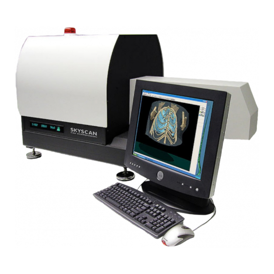
SkyScan 1076 In vivo Microtomograph
_
_
_
_
_
_
_
_
_
_
_
_
_
_
_
_
_
_
_
_
_ _
_ _
_ _
_ _
_ _
_ _
_ _
_ _
_ _
_ _
_ _
_ _
_ _
_ _
_ _
_ _
_ _
_ _
_ _
_ _
-
-
-
-
-
-
-
-
-
-
-
-
-
-
-
-
-
-
-
-
-
-
-
-
-
-
-
-
-
-
-
-
-
-
- -
- -
- -
- -
- -
- -
- -
- -
- -
- -
- -
- -
- -
- -
- -
- -
- -
- -
- -
- -
- -
- -
- -
- -
- -
- -
- -
- -
- -
- -
- -
- -
- -
Instruction Manual
_
_
_
_
_
_
_
_
_
_
_
_
_
_
_
_
_
_
_
_
_ _
_ _
_ _
_ _
_ _
_ _
_ _
_ _
_ _
_ _
_ _
_ _
_ _
_ _
_ _
_ _
_ _
_ _
_ _
_ _
-
-
-
-
-
-
-
-
-
-
-
-
-
-
-
-
-
-
-
-
-
-
-
-
-
-
-
-
-
-
-
-
-
- -
- -
- -
- -
- -
- -
- -
- -
- -
- -
- -
- -
- -
- -
- -
- -
- -
- -
- -
- -
- -
- -
- -
- -
- -
- -
- -
- -
- -
- -
- -
- -
- -
- -
_
_
_
_
_
_
_
_
_
_
_
_
_
_
_ _
_ _
_ _
_ _
_ _
_ _
_ _
_ _
_ _
_ _
_ _
_ _
_ _
_
-
-
-
-
-
-
-
-
-
-
-
-
-
-
-
-
-
-
-
-
-
-
-
-
-
- -
- -
- -
- -
- -
- -
- -
- -
- -
- -
- -
- -
- -
- -
- -
- -
- -
- -
- -
- -
- -
- -
- -
- -
-
1
Advertisement
Table of Contents

Subscribe to Our Youtube Channel
Summary of Contents for SkyScan 1076
- Page 1 SkyScan 1076 In vivo Microtomograph © SkyScan 2002 Manufactured by SkyScan n.v. Vluchtenburgstraat 3, 2630 Aartselaar Belgium Instruction Manual...
-
Page 2: Table Of Contents
1.3 Reconstruction to image................9 1.3.1 Acquisition, creation of acquisition data ..........9 1.3.2 Start of the reconstruction..............9 1.3.3 Cross-section to image ..............10 2 SKYSCAN 1076 SYSTEM OVERVIEW............11 3 INSTALLATION PROCEDURE..............14 3.1 Connections ................... 15 3.2 Power supply..................16 3.3 Starting up Skyscan1076 ............... - Page 3 SkyScan 1076 In vivo Microtomograph 5.3.6 Alignment..................46 5.3.7 View ....................46 5.3.7.1 Toolbar command ..............46 5.3.7.2 Status Bar command..............47 5.3.7.3. Zoom In ..................47 5.3.7.4 Zoom Out .................. 48 5.3.8 About in-vivo Micro-CT ..............48 5.4 Help ......................48...
-
Page 4: Introduction In X-Ray Microscopy And Microtomography
Computerised x-ray microscopy and microtomography now gives possibilities to improve the spatial resolution by seven to eight orders in the volume terms. The system "SkyScan 1076" allows to reach a spatial resolution of 15 µm corresponding to near 3x10 cubic mm voxel size. -
Page 5: Basis Principles Of Microtomogaphy
SkyScan 1076 In vivo Microtomograph 1.2 Basis principles of microtomogaphy Any x-ray shadow image is corresponding to a two-dimensional projection from the three-dimensional object. In the simplest case, we can describe it as a parallel x-ray illumination. In this approximation, each point on the shadow image contains the integration of absorption information inside the three- dimensional object in the corresponding partial x-ray beam. - Page 6 SkyScan 1076 In vivo Microtomograph Now let's rotate our object and repeat this operation. In each new rotation position of the object, we will add to the area of reconstruction the lines of possible object positions corresponding to position of shadow. This operation is named "back-projection".
- Page 7 SkyScan 1076 In vivo Microtomograph The same algorithm can produce the cross-section image not only from the single point object. Any real object can be represented as a big number of separate absorption voxels and linear absorption in any x-ray beam is corresponding to the sum of all absorption from all voxels inside this beam.
- Page 8 SkyScan 1076 In vivo Microtomograph In the case of x-ray acquisition, the image contains information about the intensity reduction inside the three-dimensional object. Because the x-ray absorption is corresponding to exponential law, we can restore the linear absorption information from the shadow image by logarithmisation.
-
Page 9: Reconstruction To Image
SkyScan 1076 In vivo Microtomograph 1.3 Reconstruction to image 1.3.1 Acquisition, creation of acquisition data During the acquisition the source-detector pair will rotate over 180 degrees. At each position the shadow image or transmission image will be acquired. Cone beam acquisitions saves all of these projection images as 16 bit TIF files. -
Page 10: Cross-Section To Image
Reconstructed array will be shown as a half-tone image of cross- section with linear conversion to 256-grades of gray inside selected density interval. In the Skyscan systems, using a Windows environment, the final image generated can be exported to BMP, RAW 16Bit or TXT -files. -
Page 11: Skyscan 1076 System Overview
SkyScan 1076 In vivo Microtomograph 2 SKYSCAN 1076 SYSTEM OVERVIEW SPECIFICATIONS: 68mm(D) x 200mm(L) for rats or 35mm(D) x 200mm(L) Maximum object size for mice, 17mm(L) per single scan 20-100kV, 10W, <5um spot size (@4W), >10000h X-ray source estimated lifetime, air cooled sealed type, 4-positions... - Page 12 SkyScan 1076 In vivo Microtomograph The "SkyScan-1076" is a high- resolution low-dose X-ray scanner for in-vivo 3D-reconstruction with spatial resolution of up to 15 microns inside the small laboratory animals (rats, mice, etc.). It consists of the combination of Micro-CT system and a computer with system control software and reconstruction software.
- Page 13 SkyScan 1076 In vivo Microtomograph For "SkyScan-1076” the X-ray microfocus tube with 5 micron focal spot size operates at 20-100kV / 0-250µA. The special X-ray CCD-camera is based on 10 Megapixel (4000x2300 pixels) cooled CCD-sensor with fibre optic coupling to x-ray scintillator The X-ray shadow projections are digitised as 1000x520 to 8000x2000 pixels with 4096 brightness gradations (12 bit).
-
Page 14: Installation Procedure
Skyscan, Vluchtenburgstraat 3, Aartselaar B-2630, BELGIUM. Email: info@skyscan.be, Tel: +32 3 877 57 05, Fax: +32 3 877 57 69 Only trained people by Skyscan are authorized to do service and reparations on the scanner! For lifting and carrying, remove the four black plastic caps and put the supplied steel bars through the holes. -
Page 15: Connections
SkyScan 1076 In vivo Microtomograph 3.1 Connections To start the system one should make the connections shown in the figure below. All interconnections should be made before connection of the main power plug to 220-240 V AC or 100-130V AC. -
Page 16: Power Supply
SkyScan 1076 In vivo Microtomograph 3.2 Power supply Never connect 200-240V to a system meant for 100-130V and vice versa. Look to the label above the AC inlet (see picture on previous page). The replaceable fuse is the same for both main voltages: type T rate 5A. Before replacement of the fuse, disconnect the power cable from the system. -
Page 17: X-Ray Shutter
SkyScan 1076 In vivo Microtomograph 3.5 X-Ray Shutter Normally the X-Ray shutter is always in the closed position. The X-ray shutter is open for acquiring one X-ray image or a scout scan and during scanning. The LED’s indicate the X-Ray shutter position: The X-ray shutter is open when the red LED lights up. -
Page 18: System Control Software
SkyScan 1076 In vivo Microtomograph 4 SYSTEM CONTROL SOFTWARE The software package for system control has the same operation principles as any other Windows-based program. One can select any functions by "mouse" from the menu / submenu in the top line and change modes and parameters by "mouse pressing"... - Page 19 SkyScan 1076 In vivo Microtomograph One can change the power settings of the tube by double clicking on the “x-ray generator on” indicator: Following dialog box will be shown onto the screen: Left part of the dialog box shows information about the type of x-ray source, elapsed time (active emission) and maximum available power.
-
Page 20: Animal Holders
SkyScan 1076 In vivo Microtomograph 4.2 Animal holders There are two standard carbon-composite object beds supplied with the system. The object bed with diameter 33 mm should be used for mice and the object bed with diameter 65 mm should be used for rats. Both have the same length of 400 mm and a maximum scanning length of 200 mm. - Page 21 SkyScan 1076 In vivo Microtomograph Remove the two screws on both sides of the bed. Now it is possible to remove the bed completely. The installation of the small (33 mm) bed needs one extra support. First, one has to install this extra support (see picture).
- Page 22 SkyScan 1076 In vivo Microtomograph Fasten the small bed with two screws on both sides and put the camera back on the small bed. Close the door by hand and start system and software again. Instruction Manual...
-
Page 23: Scout Scan And X-Ray Image
SkyScan 1076 In vivo Microtomograph 4.3 Scout Scan and X-ray image After installation of mouse or rat, close the door by hand. Mouse on animal bed with physiological monitoring sensors: To start X-ray tube push X-ray button: Voltage and current will be set automatically corresponding to current Filter and Pixel size. -
Page 24: Start Scanning
SkyScan 1076 In vivo Microtomograph 4.4 Start Scanning If the position of the bed is correct and the preview image is good enough, one can start the acquisition. Start scanning Following dialog box will be displayed onto the screen: Scanning width should correspond to the size of scanning area (diameter of object bed). -
Page 25: Reconstruction
SkyScan 1076 In vivo Microtomograph 4.5 Reconstruction One can start the external Cone-beam reconstruction program from the Toolbar: Starts reconstruction program Alternatively, one can start the Cone-beam program from the shortcut on the desktop: Following dialog box allows choosing data set for sending to reconstruction. -
Page 26: Reconstruction
SkyScan 1076 In vivo Microtomograph 4.6 3D-Reconstruction To convert the reconstructed dataset (bitmap files) into a 3D-image and to calculate the internal morphological parameters one can use the external program “3D-Creator”. One can start the program from the shortcut on the desktop:... -
Page 27: Menu And Submenu Functions
SkyScan 1076 In vivo Microtomograph 5 MENU AND SUBMENU FUNCTIONS 5.1 General layout of the main menu The top menu bar in the screen gives access to all system functions. Let's consider all menu / submenu possibilities in more details. -
Page 28: Open Image Command
SkyScan 1076 In vivo Microtomograph 5.2.1 Open Image command Use this command to open an existing image file. Following “Open” dialog will appear onto the screen. The following options allow you to specify which file to open: File Name Type or select the filename you want to open. This box lists files with the extension you select in the List Files of Type box. -
Page 29: Save Image Command
SkyScan 1076 In vivo Microtomograph 5.2.2 Save Image command Use this command to save the image to the disk. The program will display the following “Save” dialog box: The following options allow you to specify the name and type of the file you're... -
Page 30: Print Image Command And Submenu
SkyScan 1076 In vivo Microtomograph 5.2.3 Print image command and submenu The Print submenu offers the following commands: -> Prints a document -> Displays the image as it would appear when printed -> Selects a printer and a printer connection ->... - Page 31 SkyScan 1076 In vivo Microtomograph Print Layout Use this command to set print layout. Following dialog window will be displayed: Print layout for scout view contains adjustments for printing position (margins) on the page and image scale as a zoom from original (corresponding to object size) format.
-
Page 32: Delete Dataset And Submenu
SkyScan 1076 In vivo Microtomograph 5.2.4 Delete Dataset and submenu Delete Dataset -> BMP-files Use this command to remove full set of reconstructed results with selected prefix from the disk. Delete Dataset -> TIFF-files Use this command to remove full set of acquired projection images with selected prefix from the disk. -
Page 33: Show Visual Image And Physiological Monitoring
SkyScan 1076 In vivo Microtomograph 5.2.6 Show Visual Image and physiological monitoring This command will start the external program for animal imaging and physiological monitoring. Visual image of the object can be shown in one of three sizes. Shortcuts Toolbar: Icon on desktop: 5.2.6.1 The Actions menu... -
Page 34: Physiological Monitoring Subsystem
SkyScan 1076 In vivo Microtomograph 5.2.6.2 Physiological monitoring subsystem Subsystem for physiological monitoring of the small laboratory animals during scanning contains following parts: The TV-camera in the animal bed produces real-time visual image. Operator can see the animal on the computer screen in any time including x-ray scanning. -
Page 35: Physiological Monitoring Window Onto The Screen
SkyScan 1076 In vivo Microtomograph Screen copy of the Skyscan-1076 software during scanning with Physiological Monitoring. Left part: scout view (up to 200mm) with selection of the scanning position. Central part: real-time x-ray image and progress indicator; Right bottom part: physiological monitoring and real-time visual image of the animal. -
Page 36: Start Scout Scan
SkyScan 1076 In vivo Microtomograph In the Actions Menu of the “LookIn” program additional settings for temperature will appear during Physiological monitoring: -> Add this number for calibration of temperature sensor. -> Multiply with this number for calibration of temperature sensor. -
Page 37: Start Scanning For Reconstruction
SkyScan 1076 In vivo Microtomograph 5.2.8 Start Scanning for Reconstruction Use this command to start scanning to acquire data for following reconstruction. The program displays the Acquisition Parameters dialog box to adjust all necessary settings. Shortcut Toolbar: Acquisition Parameters dialog box Use this command to set the acquisition parameters. -
Page 38: Remove Object
SkyScan 1076 In vivo Microtomograph Special modes button open next dialog window with parameters settings for nonstandard scanning modes. Save only central part option allow acquiring projection images for reconstruction of specific central part of the object, which can be 1/2, 1/4, 1/8 of the selected scanning width. -
Page 39: Start Reconstruction
SkyScan 1076 In vivo Microtomograph 5.2.10 Start Reconstruction Use this command to start reconstruction program. Following dialog allows choosing data set for sending to reconstruction. Shortcut Toolbar: 5.2.11 Set Object Position Use this command to set the position of the object bed inside scanning area by numerical value. -
Page 40: Exit Command
SkyScan 1076 In vivo Microtomograph 5.2.13 Exit command Use this command to close the control program for SkyScan-1076 Micro-CT instrument. You can also use the Exit command on the Actions menu. Shortcuts Mouse: Double-clicking to the left top corner of the program window is the same as choosing the Exit command. -
Page 41: The Options Menu
SkyScan 1076 In vivo Microtomograph 5.3 The Options menu -> Sets the acquisition parameters -> Adjusts the settings for the X-Ray source -> Selects corresponding physical filter -> Access to the settings for all possible acquisition modes -> Modifies geometrical settings of the scanner. - Page 42 SkyScan 1076 In vivo Microtomograph Special modes button open next dialog window with parameters settings for nonstandard scanning modes. Save only central part option allow acquiring projection images for reconstruction of specific central part of the object, which can be 1/2, 1/4, 1/8 of the selected scanning width.
-
Page 43: X-Ray Source
SkyScan 1076 In vivo Microtomograph 5.3.2 X-Ray Source Use this command to adjust settings for x-ray source. Following dialog box will be shown onto the screen: Left part of the dialog box shows information about the type of x-ray source, elapsed time (active emission) and maximum available power. -
Page 44: Scanning Modes
SkyScan 1076 In vivo Microtomograph 5.3.4 Scanning Modes This command is only accessible by pressing “Ctrl”+”Alt”+”Shift”+”s”. Pressing the password combination again will close access to this command. Use this command for access to the settings for all possible acquisition modes: Top part of the dialog window contains a table with 12 acquisition modes for 4 available filters and 3 possible pixel sizes. -
Page 45: Set-Up
SkyScan 1076 In vivo Microtomograph 5.3.5 Set-up This command is only accessible by pressing “Ctrl”+”Alt”+”Shift”+”s”. Pressing the password combination again will close access to this command. Use this command to modify geometrical settings of the scanner. ATTENTION! Wrong values in one or several parameters can cause the scanner damages, image distortions or incorrect reconstruction results. -
Page 46: Alignment
5.3.7.1 Toolbar command Use this command to display and hide the Toolbar, which includes buttons for some of the most common commands in the control program for Skyscan-1076 micro-CT scanner. A check mark appears next to the menu item when the Toolbar is displayed. -
Page 47: Status Bar Command
SkyScan 1076 In vivo Microtomograph Click Open an image. Save the active image. Print the active image. Start or stop the x-ray source. Acquire x-ray image. Access to visual and physiological monitoring program. Start scout scanning. Start data acquisition for reconstruction. -
Page 48: Zoom Out
5.3.8 About in-vivo Micro-CT Use this command to display the copyright notice and version number of your copy of the control software for the in-vivo micro-CT scanner Skyscan-1076. 5.4 Help The Help menu offers the access to the help-information available on this software package.
Need help?
Do you have a question about the 1076 and is the answer not in the manual?
Questions and answers