Advertisement
US-6
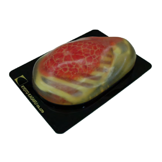
Breast Ultrasound Examination
Training Model
Unique high-fidelity ultrasound phantom facilitates effective training in basic breast
examination with your own clinical devices.
Simulated lesions embedded in as targets provide wider educational opportunities.
Any ultrasound device with a convex probe can be used for the phantom.
Anatomy in the phantom
The subcutaneous adipose the mammary gland, the galactophore, Cooper's liga-
ment, the retromammary adipose, the costae, the clavicle, the pectoralis major, the
lung and the lymph nodes at axilla.
Stable and long-life phantom materials.
" BREASTFAN "
Instruction
Manual
Contents
● Please read before training
General information
● Preparation&Training
1 Before Training
2 Training session
3 After training
Advertisement
Table of Contents

Related Manuals for Kyoto Kagaku BREASTFAN


Summary of Contents for Kyoto Kagaku BREASTFAN
- Page 1 US-6 Breast Ultrasound Examination “ BREASTFAN ” Training Model Instruction Manual Contents ● Please read before training General information ● Preparation&Training 1 Before Training 2 Training session 3 After training Unique high-fidelity ultrasound phantom facilitates effective training in basic breast examination with your own clinical devices.
- Page 2 Please read General information Before training Preparation Set Includes: Before your first use, please ensure that you have all components listed below. Ultrasound breast phantom a. b. Carrying Case c. DVD tutorial manual DOs and DON’T s DO s DON’Ts Handle with care. Never wipe the phantom or models with The materials for phantom and models are special composition thinner or organic solvent.
- Page 3 Training Simulated Targets/ Storage 2 TRAINING SESSION ●Examples of Ultrasound Images benign tumor mammary ductal ectasia ① The phantom can be scanned with real ultrasound devices. Embedded lesions lymph nodes vicious Tumor Mammary Ductal Ectasia Iymph Nodes Benign Tumor cyst Cyst Vicious Tumor 3 AFTER TRAINING ① Wipe the remaining gel completely with wet cloth. If remaining gel gets dried up on the body, it may cause scratches and tears on the phantom material.
- Page 4 Don’t mark on the phantom with pen or leave printed materials contacted on its surface. Caution Ink marks on the phantom will be irremovable. The contents of this manual are subject to change without prior notice. No part of this instruction manual may be reproduced or transmitted in any form without permission from the manufacturer.
Need help?
Do you have a question about the BREASTFAN and is the answer not in the manual?
Questions and answers