Table of Contents
Advertisement
Quick Links
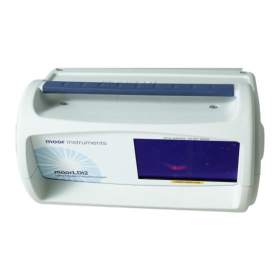
moorLDI2 USER MANUAL AND REFERENCE
RESEARCH Software Versions 5.3 Issue 4: 06-07-12
User Assistance Information
In case of difficulties which cannot be solved easily by the material within this manual, please
contact your distributor or Moor Instruments directly, quoting the instrument serial number
W o r l d w i d e
Moor Instruments Ltd
Millwey
Axminster
Devon
EX13 5HU
UK
Tel: +44 (0)1297 35715
Fax: +44 (0)1297 35716
Email:
sales@moor.co.uk
Website:
www.moor.co.uk
©2012 Moor Instruments Limited.
All rights reserved.
This document may not, in whole or in part, be copied, photocopied, reproduced, translated or be
reproduced on any electronic medium without prior consent from Moor Instruments Limited.
moorLDI is patented: UK No. 2231742, USA No. 5588437, EPC No. 0465524, Israel No. 93909,
Japan No. 3239952.
moorLDI2-VR
moorLDI2-IR
D e u t s c h l a n d
Moor Instruments GmbH
RheinAhrCampus
Südallee 2
Remagen
53424
Deutschland
Telefon: +49 (0)2642-932-232
Telefax: +49 (0)2642-932-245
Email:
sales@moorinstruments.de
Website:
www.moorinstruments.de
moorLDI2-HR
moorLDI2-HIR
U n i t e d
Moor Instruments Inc
Suite #66
501 Silverside Rd
Wilmington
DE 19809
USA
Tel: (302) 798 7470
Fax: (302) 798 7299
Email:
Website:
S t a t e s
sales@moorinc.com
www.moorinc.com
Advertisement
Table of Contents

Summary of Contents for Moor Instruments moorLDI2-VR
- Page 1 User Assistance Information In case of difficulties which cannot be solved easily by the material within this manual, please contact your distributor or Moor Instruments directly, quoting the instrument serial number W o r l d w i d e...
- Page 2 MOOR INSTRUMENTS moorLDI2 RESEARCH USER MANUAL EC DECLARATION OF CONFORMITY Moor Instruments Ltd Millwey Axminster Devon EX13 5HU England Declares that the medical device described hereafter: moorLDI2 Laser Doppler Imager is in conformity with the essential requirements and provisions of Council Directive...
-
Page 3: Table Of Contents
MOOR INSTRUMENTS moorLDI2 RESEARCH USER MANUAL TABLE OF CONTENTS SAFETY ..................................1 INTRODUCTION AND INTENDED USE ......................1 LASER SAFETY ..............................1 1.2.1 LASER SAFETY moorLDI2-VR AND moorLDI2-HR ................... 2 1.2.1.1 Training ................................2 Unauthorised Access ............................. 2 1.2.1.2 1.2.1.3 Warning Signs ............................... - Page 4 MOOR INSTRUMENTS moorLDI2 RESEARCH USER MANUAL CHANGING SCAN PARAMETERS ........................40 MOORLDI2-HR AND MOORLDI2-HIR SCAN DISTANCE AND SCAN SPEED ........... 40 MOORLDI2 SET UP .............................. 41 INTRODUCTION ..............................41 LDI2 IMAGE SCAN CONFIGURATION ....................42 MOOR 6.2.1 INTRODUCTION ............................42 6.2.2...
- Page 5 MOOR INSTRUMENTS moorLDI2 RESEARCH USER MANUAL IMAGE REVIEW ..............................65 10.1 INTRODUCTION ............................65 10.2 ....................65 UMMARY OF MAGE ROCESSING TILITIES 10.3 OPENING FILES IMAGE DISPLAY ..................... 68 10.3.1 PHOTO IMAGE DISPLAY ........................68 10.3.2 FLUX IMAGE DISPLAY ........................68 10.3.3...
- Page 6 MOOR INSTRUMENTS moorLDI2 RESEARCH USER MANUAL 15.1.1 TYPES OF IMAGE ANALYSIS ......................87 15.1.2 PIXEL VALUES ............................ 87 15.2 STATISTICS - SINGLE IMAGE SCAN ......................88 15.2.1 FILE - SAVE ............................88 15.2.2 FILE - APPEND ............................ 88 15.2.3 FILE - PRINT ............................89 15.2.4...
- Page 7 MOOR INSTRUMENTS moorLDI2 RESEARCH USER MANUAL MAINTENANCE ..............................119 20.1 LASER OUTPUT POWER ........................... 119 20.2 LASER BEAM PROPERTIES ........................119 20.3 MECHANICAL CHECKS ..........................119 20.4 OPERATION OF THE SOLENOID ACTUATED OPTICAL ATTENUATOR .......... 120 20.5 LEADS ................................120 20.6...
-
Page 8: Safety
MOOR INSTRUMENTS moorLDI2 RESEARCH USER MANUAL 1. SAFETY INTRODUCTION AND INTENDED USE The ‘Moor Instruments moorLDI2 User Manual’ reference for the following imager types: moorLDI2-VR Visible red laser Class 3R moorLDI2-IR Near infra-red laser Class 3R moorLDI2-HR High Resolution version of moorLDI2-VR Class 3R... -
Page 9: Laser Safety Moorldi2-Vr And Moorldi2-Hr
MOOR INSTRUMENTS moorLDI2 RESEARCH USER MANUAL 1.2.1 LASER SAFETY moorLDI2-VR AND moorLDI2-HR The laser light source in these moorLDI2 scanners is a Helium-Neon visible red gas laser (633nm). The maximum accessible power is 2.5mW for instruments using the gas laser. -
Page 10: Warning Signs
MOOR INSTRUMENTS moorLDI2 RESEARCH USER MANUAL 1.2.1.3 Warning Signs The entrance to areas where the moorLDI2 is to be used should be posted with appropriate warning signs: DANGER Laser Hazard No Unauthorised Entry 1.2.1.4 Safety During Operation It is not generally necessary for the patient to wear protective eyewear; however the patient must always have eye protection if the patient's face is to be scanned. -
Page 11: Laser Safety Moorldi2-Ir And Moorldi2-Hir
(660nm) target beam to be clearly seen. All laser protective eyewear should be clearly identified as being suitable for use with the moorLDI2-IR. Moor Instruments will normally supply protective eye wear that can be worn over prescription glasses. -
Page 12: Laser Safety Officer
MOOR INSTRUMENTS moorLDI2 RESEARCH USER MANUAL 1.2.2.2 Laser Safety Officer For installations where Class 3R laser products emitting energy outside of the 400nm to 700nm wavelength ranges are operated, a Laser Safety Officer should be appointed. It is the Laser Safety Officer's responsibility to review the following precautions and designate the appropriate controls to be implemented. -
Page 13: Key Control
MOOR INSTRUMENTS moorLDI2 RESEARCH USER MANUAL being scanned such that if the laser light beam is missing the body it is stopped by the screen. A dark cloth screen is recommended. If a suitable screen is not available the body should be scanned with a wall as background. -
Page 14: Beam Delivery System
MOOR INSTRUMENTS moorLDI2 RESEARCH USER MANUAL BEAM DELIVERY SYSTEM The laser light source assembly consists of a visible red laser diode with a collimating lens to produce a beam of laser light (see Figure 1, below) A solenoid operated attenuator mounted in front of the laser aperture of the assembly can be switched between a full power working beam (<2.5mW visible red) and an attenuated beam (<200µW). -
Page 15: Laser Warning Labels
MOOR INSTRUMENTS moorLDI2 RESEARCH USER MANUAL LASER WARNING LABELS Location: Front panel adjacent to "LASER READY" indicator Location: Scanner head rear panel CAUTION - CLASS 3R LASER RADIATION (for Visible Red System Class 3R) WHEN OPEN AVOID DIRECT EYE EXPOSURE... -
Page 16: Electrical Safety Labels
MOOR INSTRUMENTS moorLDI2 RESEARCH USER MANUAL ELECTRICAL SAFETY LABELS WARNING ISOLATE MACHINE BEFORE REMOVING COVERS Location: Control box rear panel ATTENTION ISOLER MACHINE AVANT D'ENLEVER COUVERCLES ACHTUNG MASCINE ISOLIEREN BEVOR ENTFERNUNG DER DECKEL AVISO AISLE LA MAQUINA ANTES DE QUITAR... -
Page 17: Environmental Conditions
Do not attempt to clean the optical window unless you have been instructed how to do so and have been authorised to do so - the surface is easily scratched. If scratched it will have to be replaced. Do consult the responsible person for the moorLDI2 in your establishment or a Moor Instruments representative if you need advice. -
Page 18: System Description
100 - 120V 1 A (T) 50 - 60 Hz AISLE LA MAQUINA ANTES DE QUITAR 50 VA Made in England by Moor Instruments Ltd Millwey, Axminster, Devon, EX13 5HU 220 - 240V 500 mA (T) ATTENZIONE Tel: +44 (0)1297 35715... - Page 19 100 - 120V 1 A (T) 50 - 60 Hz AISLE LA MAQUINA ANTES DE QUITAR 50 VA Made in England by Moor Instruments Ltd Millwey, Axminster, Devon, EX13 5HU 220 - 240V 500 mA (T) ATTENZIONE Tel: +44 (0)1297 35715...
- Page 20 MOOR INSTRUMENTS moorLDI2 RESEARCH USER MANUAL Figures 2D and 2E show the layout of the front panel and back panel labels of the moorLDI2 single visible wavelength scanner units moorLDI2-VR, moorLDI2-HR Warning LED’s: 1, Power; 2, Laser ready, 3, The Laser Aperture , is indicated and shown here schematically by the rectangle above.
- Page 21 MOOR INSTRUMENTS moorLDI2 RESEARCH USER MANUAL Figures 2F and 2G show the layout of the front panel and back panel labels of the moorLDI2 single infrared wavelength scanner units. moorLDI2-IR and moorLDI2-HIR FIGURE 2F - FRONT PANEL OF THE moorLDI2 INFRARED SCANNER UNIT...
-
Page 22: Moorldi2 Unpacking And Assembly
MOOR INSTRUMENTS moorLDI2 RESEARCH USER MANUAL moorLDI2 UNPACKING AND ASSEMBLY The moorLDI2 will initially be installed by a Moor Instruments engineer or approved representative. The following instructions should be followed for subsequent installations. The various stages of assembly prior to operating the moorLDI2 are: a. -
Page 23: Basic Stand (Moorldi-Bs1)
MOOR INSTRUMENTS moorLDI2 RESEARCH USER MANUAL The Transverse Arm This passes through the winder box. First remove the handle from the transverse arm and loosen the knobs on the side of the winder box. Pass the transverse arm, from the front, through the winder box (the longest forks of the H frame are at the front). -
Page 24: Unpacking The Scan Head
1 off moorLDI2 25-way scan head-to-controller cable (Basic stand and Desktop stand only) 1 off moorLDI2 scan head-to-PPC framegrabber video cable 1 off framegrabber card (if a PPC Kit was supplied by Moor Instruments, the framegrabber card will already be installed and tested in the PC supplied) -
Page 25: Installation Of Firewire Card (Firewire Camera Model)
Please note not all laptop computers with Firewire interface work with the Firewire camera, therefore only a laptop supplied by Moor Instruments can be guaranteed to work. To install the card in a desktop or panel PC follow these steps: Switch off your computer and all peripheral devices. -
Page 26: Cable Connections
MOOR INSTRUMENTS moorLDI2 RESEARCH USER MANUAL 3.5 CABLE CONNECTIONS If a clinical mobile stand has been supplied, refer directly to Section 3.1.1 N.B. the mains supply to all equipment should be turned OFF until all connections have been made. Connect cables as shown schematically below: 3.5.1 CABLING... - Page 27 MOOR INSTRUMENTS moorLDI2 RESEARCH USER MANUAL Power RS232 lead 8 Way 25 Way cable cable screened cable FIGURE 3B – BACK OF THE CONTROL BOX SHOWING CABLES AND THE CORRESPONDING CONNECTIONS (ANALOGUE CAMERA MODEL)
- Page 28 MOOR INSTRUMENTS moorLDI2 RESEARCH USER MANUAL 8 way 25 way screened Video cable cable connector FIGURE 3C – BACK OF THE SCAN HEAD AND CONNECTING CABLES (ANALOGUE CAMERA MODEL) Power cable RS232 lead Video cable FIGURE 3D – PC CONNECTION CABLES (ANALOGUE CAMERA MODEL)
- Page 29 If you are unable to identify the connector or if it has 25 pins rather than 9 please contact Moor Instruments for further advice. Secure the connectors with the screw fixings provided which should be finger tight.
- Page 30 MOOR INSTRUMENTS moorLDI2 RESEARCH USER MANUAL Firewire Camera Model: 25 WAY SCREENED CABLE PORT 1 25 WAY 'D' TYPE SCANNER 8 WAY SCREENED CABLE CONTROL 8 WAY PORT 2 HEAD Circular TYPE VIDEO LINK PHONO Video SOCKET IEEE 1394 RS232...
- Page 31 MOOR INSTRUMENTS moorLDI2 RESEARCH USER MANUAL 8 way 25 way screened Video cable cable connector FIGURE 3G – BACK OF THE SCAN HEAD AND CONNECTING CABLES (FIREWIRE CAMERA MODEL) Power cable RS232 lead Firewire Cable FIGURE 3H – PC CONNECTION CABLES (FIREWIRE CAMERA MODEL)
- Page 32 If you are unable to identify the connector or if it has 25 pins rather than 9 please contact Moor Instruments for further advice. Secure the connectors with the screw fixings provided which should be finger tight.
-
Page 33: Mobile Stand And Cabling
MOOR INSTRUMENTS moorLDI2 RESEARCH USER MANUAL 3.5.2 MOBILE STAND AND CABLING Analogue Camera Model: Three of the cables (8-way, 25-way and video) for use with a moorLDI2-MS2 mobile stand are in a corrugated sleeve. This wiring loom is shipped in the THB transformer housing box. At one end the leads are short and of similar length (Figure 3C). -
Page 34: The Computer And Software Installation
- Safety - Part 1: General requirements). Note that generally any commercially available PC will meet this standard. If the PC is supplied by Moor Instruments it will meet this standard and meet or exceed the minimum recommended specification detailed below. - Page 35 MOOR INSTRUMENTS moorLDI2 RESEARCH USER MANUAL To install the drivers and the moorLDI2 V5.3 software follow these steps: Make sure that the IEEE Firewire cable is not connected to the PC before installing software and turn off the moorLDI2. Insert the software installation CD-ROM disc into your CD-ROM drive.
- Page 36 MOOR INSTRUMENTS moorLDI2 RESEARCH USER MANUAL The driver will begin to install. A warning that the driver has not passed Windows Logo testing may be displayed. If this happens, click Continue Anyway . For Windows Vista click on the “ Install this driver software anyway”.
- Page 37 MOOR INSTRUMENTS moorLDI2 RESEARCH USER MANUAL Driver installation is complete when the following window is displayed. Click Finish to complete driver installation. To finish installing the software click on Finish when the following window is displayed:...
- Page 38 MOOR INSTRUMENTS moorLDI2 RESEARCH USER MANUAL Connect the RS232 or USB cable to the PC and the system is ready for use. The above installation is designed for use with the new Firewire camera model, if you are using the analogue camera with the F6 card and the F6 driver has not been installed on the PC before, you will need to install the driver separately after the above installation.
-
Page 39: Before First Use
• Check that the distance measurement is accurate to ± 5cm at 80cm scan distance If you suspect your system may have been damaged, or if your system fails the calibration or distance checks, please contact your distributor or Moor Instruments directly, quoting the instrument serial number... -
Page 40: The Laser Doppler Technique
MOOR INSTRUMENTS moorLDI2 RESEARCH USER MANUAL THE LASER DOPPLER TECHNIQUE INTRODUCTION The laser Doppler technique was first applied to monitoring tissue blood flow by Stern et al in 1975. Since then it has been applied to most branches of medicine and physiology. -
Page 41: Elements Of The Moorldi2
4.4.1 THE LASER SOURCE The laser wavelength, accessible power and laser classification depend on the moorLDI2 version. moorLDI2-VR and moorLDI2-HR Class 3R visible laser. Either a Helium-Neon gas laser wavelength 633nm, maximum accessible power 2.5mW. Maximum scan area with large scan setting approximately 60 x 60 cm. -
Page 42: The Laser Doppler Processing Algorithm
MOOR INSTRUMENTS moorLDI2 RESEARCH USER MANUAL THE LASER DOPPLER PROCESSING ALGORITHM After detection and amplification the signals are digitised and processed to yield flux (proportional to blood flow) and conc (proportional to the concentration of moving blood cells). The algorithms used to compute these parameters are: ω... - Page 43 MOOR INSTRUMENTS moorLDI2 RESEARCH USER MANUAL Flow Units It is generally agreed by researchers and manufacturers that because of the nature of the flow in capillaries and connecting small blood vessels and the effect of varying skin colour and structure, it is not generally appropriate to use absolute flow units such as ml/min/100gm.
-
Page 44: Introduction To Moorldi2 Software
MOOR INSTRUMENTS moorLDI2 RESEARCH USER MANUAL INTRODUCTION TO moorLDI2 SOFTWARE Once loaded, the moorLDI2 V5.3 software presents the following options MEASUREMENT To obtain Laser Doppler Measurements (scan, repeat scan, repeat line and single point measurement) IMAGE REVIEW Image processing and analysis TRACE REVIEW To review single point measurements. -
Page 45: Overview
MOOR INSTRUMENTS moorLDI2 RESEARCH USER MANUAL OVERVIEW The MEASUREMENT module has utilities required for: - obtaining LDI images and recording patient information - recording images- single and repeat scans - recording line images - recording colour video images - controlling the imager function... -
Page 46: Getting Started With The Moorldi2
MOOR INSTRUMENTS moorLDI2 RESEARCH USER MANUAL GETTING STARTED WITH the moorLDI2 For some early versions of the moorLDI instrument it is necessary to input a registration number and the serial number of the scanner. For these instruments an error message will be displayed when the green start button is clicked (see item 8 below). -
Page 47: Changing Scan Parameters
MOOR INSTRUMENTS moorLDI2 RESEARCH USER MANUAL You will be prompted to either manually enter the scan head to tissue distance or confirm the automatic distance measurement itself. Click on the green flag start icon Note you can pause or stop the scan at any time). -
Page 48: Moorldi2 Set Up
MOOR INSTRUMENTS moorLDI2 RESEARCH USER MANUAL MOORLDI2 SET UP This section describes how to set up the moorLDI2 system for a specific measurement. INTRODUCTION The measurement module presents the following display header when a scan window has been opened, until then only icons 2 to 8 are shown and are active. -
Page 49: Moorldi2 Image Scan Configuration
MOOR INSTRUMENTS moorLDI2 RESEARCH USER MANUAL moorLDI2 IMAGE SCAN CONFIGURATION 6.2.1 INTRODUCTION Single Image Acquisition should be used when only one image is required. If several images are to be recorded at fixed time intervals the repeat scan function (described later in this manual Section 8 ) can be chosen. -
Page 50: Scan Size, Area And Resolution
MOOR INSTRUMENTS moorLDI2 RESEARCH USER MANUAL The maximum scan area is also displayed in this window. If the size displayed is not large enough either: - increase the moorLDI -to-tissue distance, - click on Large to double the area which can be scanned compared to the Normal setting. -
Page 51: Video And Distance
MOOR INSTRUMENTS moorLDI2 RESEARCH USER MANUAL To view a scan region on the tissue: click Mark in LD Photo / Mark Beam or press the M key, to start continuous marking. click Abort or press the A key to end the continuous marking.. -
Page 52: Mark Beam
MOOR INSTRUMENTS moorLDI2 RESEARCH USER MANUAL When changing scan size between Large and Normal , a zoom change will be observed. 6.3.2.1 Mark Beam The laser beam is visible in the Live Video Image screen. The beam position can be changed, in Beam Movement , by changing the values in the X and Y boxes, or clicking the left/right arrows. -
Page 53: Repeat And Line Scan Setup
MOOR INSTRUMENTS moorLDI2 RESEARCH USER MANUAL This is particularly important for applications where regular changes of distance are made (e.g. burn assessments). Note that should the wrong distance be used during measurement it is possible to change the distance at the data review stage. -
Page 54: Line Scan Setup
MOOR INSTRUMENTS moorLDI2 RESEARCH USER MANUAL If the Repeat Interval is set to 0:0:0, a fast repeat scan mode will be selected where the solenoid will not be turned off between each succesive scan. This ensures that the time between the finish of one scan and the start of the next is a minimum. -
Page 55: Normalisation
MOOR INSTRUMENTS moorLDI2 RESEARCH USER MANUAL Automatic Set is selected by default. The upper cutoff frequency will be 15KHz and the lower cutoff will be automically selected to suit the scan speed. (20Hz for 50ms/pixel, 100Hz for 10 ms/pixel and 250Hz for 4ms/pixel.) -
Page 56: Auto Gain
MOOR INSTRUMENTS moorLDI2 RESEARCH USER MANUAL the limit of the 12 bit A/D convertor). For distances of about 40 to 60 cm the Factory Setup gains of 2, 2, 2 will probably be ok for most measurements. Gain settings can be entered in the DC, Flux and Conc boxes or the gains can be set automatically through Gain Setup. -
Page 57: Reading (Ru)
MOOR INSTRUMENTS moorLDI2 RESEARCH USER MANUAL The DC gain should be reduced: in this example from 2 to the 0. When moist tissue is being scanned, it is important to avoid scanning perpendicular to the tissue by angling the scan head (about 15degrees) to avoid direct laser reflection. -
Page 58: Single Point Measurements
MOOR INSTRUMENTS moorLDI2 RESEARCH USER MANUAL 6.3.5 SINGLE POINT MEASUREMENTS The Single Point Measurement (SPM) function enables real time monitoring of laser Doppler flux and conc at a point on the tissue surface at relatively high data rates (upto 40Hz). -
Page 59: Setup: Preference Setup
MOOR INSTRUMENTS moorLDI2 RESEARCH USER MANUAL 6.4 Setup: Preference Setup Opens the Preference Setup dialogue box as shown below: Save the last DC and Flux Palette settings when Exit the Software: Select this option to remember the Palette settings when the software is closed. - Page 60 MOOR INSTRUMENTS moorLDI2 RESEARCH USER MANUAL Select this option to delete the temporary files (saved in the C: root directory) when the corresponding measurement window is closed. Show Repeat Interval Confirmation Message at the Beginning of Repeat Scan: Select this option to enable the warning message when the selected interval is shorter than the estimated scan (one image) time.
-
Page 61: Single Image Scan
MOOR INSTRUMENTS moorLDI2 RESEARCH USER MANUAL SINGLE IMAGE SCAN TO START After setting up the scan window using Setup and configuring the system (or loading a previously defined configuration file) it is possible to proceed to imaging. Click on the single scan icon... -
Page 62: Saving An Image Scan
MOOR INSTRUMENTS moorLDI2 RESEARCH USER MANUAL Once the distance has been entered, click OK to start scanning. SAVING AN IMAGE SCAN On completion of an image scan it can be saved by clicking on the save icon or by selecting File... - Page 63 MOOR INSTRUMENTS moorLDI2 RESEARCH USER MANUAL Toggle between full size and actual size image display. Toggle between PU (perfusion units) and RU (relative units). To open palette window for changing image display range. These actions are self evident except for 6 and 7 which are described further in Sections 9.9 and 9.10,...
-
Page 64: Repeat And Line Scan Measurement
MOOR INSTRUMENTS moorLDI2 RESEARCH USER MANUAL REPEAT AND LINE SCAN MEASUREMENT INTRODUCTION There are many situations where it is an advantage to take successive measurements of the same tissue site to observe a response evolve in timed sequence. The minimum time between successive measurements is determined by the size and resolution of the images to be taken. -
Page 65: Repeat Scan Start
MOOR INSTRUMENTS moorLDI2 RESEARCH USER MANUAL REPEAT SCAN START After setting up the scan window, system configuration and repeat scan configuration (or loading a previously defined configuration file) you can proceed to Repeat Scan measurement. Click on the Repeat Scan icon... -
Page 66: Repeat Scan Display During Measurement
MOOR INSTRUMENTS moorLDI2 RESEARCH USER MANUAL To start click on the green flag. REPEAT SCAN DISPLAY DURING MEASUREMENT If a small area has been selected for a repeat scan it can be displayed either as the small portion it occupies in the image window or occupying the full scan window: click on the expand icon to toggle these options (see Section 6.3, icon 1). -
Page 67: Repeat Line Scan Start
MOOR INSTRUMENTS moorLDI2 RESEARCH USER MANUAL REPEAT LINE SCAN START Once the repeat line has been defined, click on to open a window for repeat line measurement. Click the green arrow to start. Save the finished measurement in the same way as you would a single scan. -
Page 68: Single Point Measurement (Spm)
MOOR INSTRUMENTS moorLDI2 RESEARCH USER MANUAL SINGLE POINT MEASUREMENT (SPM) INTRODUCTION The Single Point Measurement (SPM) function enables real time monitoring of laser Doppler flux and conc at a point on the tissue surface. POSITIONING OF BEAM FOR SPM To measure at the point of interest it is first necessary either to move the tissue to the beam position or to move the beam to the position required. -
Page 69: Distance - Moorldi2-To-Tissue
MOOR INSTRUMENTS moorLDI2 RESEARCH USER MANUAL On entering this window 14 new icons appear: Start: to start a single point measurement. Stop: to stop a single point measurement. Pause/continue: to interrupt a single point measurement. To expand the time scale. -
Page 70: Autogain
MOOR INSTRUMENTS moorLDI2 RESEARCH USER MANUAL Select using the Scanner Setup icon SetUp Scanner Setup Click the right-most tab to configure the single point measurement. Parameters that can be altered include; Monitor Duration: free run/preset time (Preset time in Hours: Minutes: Seconds. -
Page 71: Single Point Measurement - File Store
MOOR INSTRUMENTS moorLDI2 RESEARCH USER MANUAL click on the green flag start icon You can expect to see pulsatile flow from a warm finger – see the example below. To stop, click on the stop icon To pause, click on the pause icon 9.10... -
Page 72: Image Review
MOOR INSTRUMENTS moorLDI2 RESEARCH USER MANUAL IMAGE REVIEW 10.1 INTRODUCTION The image processing utilities enhance the displayed image for various applications. In this section are described image display utilities. Image processing is described in Section 10 and Analysis in Section 14. - Page 73 MOOR INSTRUMENTS moorLDI2 RESEARCH USER MANUAL Other icons become active when an image file is opened. These are:- 2 3 4 5 6 8 9 10 11 12 13 14 15 16 17 18 Save Current File with different name...
- Page 74 MOOR INSTRUMENTS moorLDI2 RESEARCH USER MANUAL Reload - reload original unprocessed image - as 8 above Interpolate - as 10 above Normalise - alter image normalisation – note default is division by DC Rotate - Rotate Left/Rotate Right Flip - FlipHorizontal/Flip Vertical Flux:Adjust Settings - change background level.
-
Page 75: Opening Files And Image Display
MOOR INSTRUMENTS moorLDI2 RESEARCH USER MANUAL 10.3 OPENING FILES and IMAGE DISPLAY Open a file by clicking the icon or select File , Open . By default only *.flx file names for single images are displayed in the directory window. Other types (repeat scan/ repeat line/ JPG video and BMP video files) can be displayed by selection from the window below this. -
Page 76: Flux And Photo Image Display
MOOR INSTRUMENTS moorLDI2 RESEARCH USER MANUAL 10.3.3 FLUX AND PHOTO IMAGE DISPLAY To display both flux and photo images click on the split camera/hand icon . To obtain the correct aspect ratio of the images, see Aspect below. 10.4 VIEW 10.4.1 ASPECT... -
Page 77: Palette
MOOR INSTRUMENTS moorLDI2 RESEARCH USER MANUAL The moorLDI2 imager is calibrated using a motility standard consisting of polystyrene microspheres in water. Calibration is done is done during the final testing before it leaves the factory. Calibration can be checked, and the imager recalibrated if necessary, by the user. -
Page 78: Image Information
MOOR INSTRUMENTS moorLDI2 RESEARCH USER MANUAL 10.4.4 IMAGE INFORMATION All the configuration parameters and operator entered data is stored with the image and can be recalled by clicking the information icon... -
Page 79: Paste Flux Image
MOOR INSTRUMENTS moorLDI2 RESEARCH USER MANUAL 10.4.5 PASTE FLUX IMAGE One or more (uptp 5) rectagular ROIs on the Flux image can be pasted on to the Photo Image. -
Page 80: Zoom In
MOOR INSTRUMENTS moorLDI2 RESEARCH USER MANUAL 10.4.6 ZOOM IN To enlarge a rectangular region of an image it is first necessary to define the rectangular region of interest. Zoom the ROI by clicking on the magnifying glass icon with the '+' sign. The zoomed image is opened in a fresh Window automatically. -
Page 81: Image Processing
MOOR INSTRUMENTS moorLDI2 RESEARCH USER MANUAL 11. IMAGE PROCESSING 11.1 RELOAD This function is to re-load the original file ie the unprocessed image. 11.2 INTERPOLATE Low resolution or small images can be enhanced to increase the apparent resolution by interposing mean values between adjacent pixels. -
Page 82: Rotate Or Flip
MOOR INSTRUMENTS moorLDI2 RESEARCH USER MANUAL 11.4 ROTATE or FLIP The image can be rotated left (anti clockwise) or right (clockwise) by 90 o each time the operation is applied. Images can also be flipped horizontally or vertically to give a mirror image of its former state. -
Page 83: Smooth
MOOR INSTRUMENTS moorLDI2 RESEARCH USER MANUAL 11.6 SMOOTH Some features of flux and photo images can be easier to see if the images are smoothed one or more times. Smoothing is an averaging operation using the pixel value and the values of the pixels nearest neighbours. -
Page 84: Add Constant
MOOR INSTRUMENTS moorLDI2 RESEARCH USER MANUAL 11.9 CONSTANT (+,-,x,/) 11.9.1 ADD CONSTANT Add a constant to the flux image. 11.9.2 SUBTRACT CONSTANT Subtract a constant from the flux image. 11.9.3 MULTIPLY CONSTANT Enables rescaling of an image. An example of its use might be to set baseline images to similar values so that subsequent responses can be compared. -
Page 85: Threshold/ Cut
MOOR INSTRUMENTS moorLDI2 RESEARCH USER MANUAL 11.10 THRESHOLD/ CUT The Threshold function is used to discount pixels either above or below user set flux or DC values. Threshold can be applied to both Blood Flow images and LD Photo images. This can be used to assess the extent of flare removing pixels that are saturated on either flux or photo images and displaying them as background grey. -
Page 86: File
MOOR INSTRUMENTS moorLDI2 RESEARCH USER MANUAL FILE 12.1 SAVED IMAGE FILE FORMATS After original files have been processed you might wish to save the file in its processed form (i.e. smoothed, interpolated, palette change etc). Save As This operation willsave the active image in a moorLDI format. These images can only be displayed using the moorLDI software. -
Page 87: Redefine Ldi File
MOOR INSTRUMENTS moorLDI2 RESEARCH USER MANUAL 12.2 REDEFINE LDI FILE Save Active ROI image: if there are rectangle ROIs defined, images within the active rectangle ROI will be saved into a new file. Combine Single Image Files: combine single image files into one repeat image file. The single images must have the same LDI type, Scan Size, Scan Range and Resolutions. -
Page 88: Print Report
MOOR INSTRUMENTS moorLDI2 RESEARCH USER MANUAL 12.3 PRINT REPORT A report printing function is avaliable: If you select: File or click the icon Print Report Images will be printed together with information relating to the patient , operator , file name, scan setup conditions and comments. -
Page 89: Repeat Scan Processing
MOOR INSTRUMENTS moorLDI2 RESEARCH USER MANUAL REPEAT SCAN PROCESSING 13.1 INTRODUCTION All the utilities for processing and analysing single images can also be used on repeat scan images. A powerful function of the repeat scan utility is the simultaneous processing of all images. -
Page 90: Separate Image Display
MOOR INSTRUMENTS moorLDI2 RESEARCH USER MANUAL 13.3 SEPARATE IMAGE DISPLAY In addition to all the display and image processing options for single images (described in Section 10) the repeat images can also be displayed individually: by clicking on the 9-square icon... -
Page 91: Region Of Interest
MOOR INSTRUMENTS moorLDI2 RESEARCH USER MANUAL REGION OF INTEREST 14.1 INTRODUCTION The simplest form of image analysis is to obtain the flux and dc values at an image position. These are shown at the bottom of the screen when the cursor is placed at any position on either the flux or the photo image. -
Page 92: Defining Roi' - General
MOOR INSTRUMENTS moorLDI2 RESEARCH USER MANUAL 14.3 DEFINING ROI’s - GENERAL Click on either a rectangular, circular, polygon or line followed by the Add ROI button or the Insert key , and the choosen ROI will appear on the Flux and Photo images, if in single image mode, or on all the Flux images if repeat scans are being displayed. -
Page 93: Circular Roi
MOOR INSTRUMENTS moorLDI2 RESEARCH USER MANUAL When the ROI is complete the statistics or a Histogram can be constructed for the flux values within. 14.6 CIRCULAR ROI Click on the Circle ROI icon and press insert key to create a circular ROI. -
Page 94: Image Analysis
MOOR INSTRUMENTS moorLDI2 RESEARCH USER MANUAL IMAGE ANALYSIS 15.1 INTRODUCTION To analyse a either a whole image or sections of an image or equivalent parts of each image within a repeat scan set, it is first necessary to load the image(s) onto screen:... -
Page 95: Statistics - Single Image Scan
MOOR INSTRUMENTS moorLDI2 RESEARCH USER MANUAL 15.2 STATISTICS - SINGLE IMAGE SCAN After defining a region of interest click on the statistic icon An information window will be displayed similar to that shown below. 15.2.1 FILE - SAVE The statistical information can be saved to an ASCII file (*.sta) by selecting F ile, S ave. A file directory should be selected and filename given in the Save As window which appears. -
Page 96: File - Print
MOOR INSTRUMENTS moorLDI2 RESEARCH USER MANUAL 15.2.3 FILE - PRINT Select F ile, P rint to print out the statistics window. 15.2.4 FILE – EXIT Select E xit to remove the statistics window. Users of Windows ™ 95 can click on the X at the top right hand corner of the window to close. -
Page 97: Histogram Limits
MOOR INSTRUMENTS moorLDI2 RESEARCH USER MANUAL Black and Grey mode when this option is selected, bin number and lower/upper limits are editable by changing the upper and lower limits of thje histogram. Alternative black and grey colours are used to plot the histogram bars. -
Page 98: Bin Number
MOOR INSTRUMENTS moorLDI2 RESEARCH USER MANUAL Changing the lower limit will make the first bin start at the new lower limit. The graph will remain with the origin at (0, 0). The amplitude of the histogram (y-axis scale) can be increased using the + box or decreased using the –... -
Page 99: Profile
MOOR INSTRUMENTS moorLDI2 RESEARCH USER MANUAL 15.4 PROFILE 15.4.1 INSERTING PROFILE LINES The Profile analysis can only be selected after a line has been defined across an image. To achieve an averaging affect a wide line (in the example below it is 10 pixels wide) can be defined from the profile window. -
Page 100: Thickness Of Profile Line
MOOR INSTRUMENTS moorLDI2 RESEARCH USER MANUAL 15.4.3 THICKNESS OF PROFILE LINE Enter the thickness (usually between 2 to 10 ) and click the Plot box to see the new profile graphs. The default thickness of the profile line is one pixel. This often results in a noisy-looking profile and the appearance can be improved by increasing the thickness of the line to include pixels adjacent to the line. - Page 101 MOOR INSTRUMENTS moorLDI2 RESEARCH USER MANUAL moorLDI Image Profile (WRFELBI3.FLX) ============================================================== Line No Thickness Average-Dir Length(cm) Flux Profile (Perfusion Unit): Line1 1107 1035 1057 1006 1165 1156 1354 1144 1396 1186 1401 1221 1221 1181 1338 1112 1267 1032 1041...
-
Page 102: Repeat Image Analysis
MOOR INSTRUMENTS moorLDI2 RESEARCH USER MANUAL REPEAT IMAGE ANALYSIS 16.1 INTRODUCTION The analyses available for single image processing are also available for repeat scan images. All repeat images are processed and analysed simultaneously. To open a repeat image file click on icon and select from ‘files of type’... -
Page 103: Statistics - Repeat Scan
MOOR INSTRUMENTS moorLDI2 RESEARCH USER MANUAL This feature can be useful for observing more clearly responses to a stimulus by subtracting a baseline image from all other images. Enter the number of the image to be subtracted. You can also choose to average the first several repeat images then use this averaged image as the base image for subtraction or select a single image file. -
Page 104: Histogram: Repeat Scan
MOOR INSTRUMENTS moorLDI2 RESEARCH USER MANUAL To display the statistics numerically select Numerical Table from the menu. 16.1.3 HISTOGRAM: REPEAT SCAN After defining Regions of Interest (ROI, see Section14) for the repeat images histograms (section 15.3) of the flux levels can be constructed. -
Page 105: Profile: Repeat Scan
MOOR INSTRUMENTS moorLDI2 RESEARCH USER MANUAL 16.1.4 PROFILE: REPEAT SCAN Profile lines can be placed on the repeat images by clicking the profile icon and the insert icon Profiles are displayed by clicking the ‘open profile window’ icon In the example the profiles for line 2 are plotted. The controls Palette, Auto, Y Inc, Y Dec and... -
Page 106: Saving Results Of Repeat Images
MOOR INSTRUMENTS moorLDI2 RESEARCH USER MANUAL 16.2 SAVING RESULTS OF REPEAT IMAGES All results from repeat image analysis can be stored into ASCII format files. These can be imported into spreadsheet packages for further processing or inspected visually using Windows ™ Notepad. - Page 107 MOOR INSTRUMENTS moorLDI2 RESEARCH USER MANUAL A File menu is accessible at the top of the Line Image Trace window. The trace can be saved/appended into a text file. The trace displayed can be printed with or without the grid.
-
Page 108: Trace Review (Single Point Measurements)
MOOR INSTRUMENTS moorLDI2 RESEARCH USER MANUAL TRACE REVIEW (SINGLE POINT MEASUREMENTS) 17.1 INTRODUCTION Traces of the blood flow flux and conc values obtained by single point measurement are reviewed by a program separate from the Image Review program. To run this program click on the Trace Review (single point measurement) icon 17.2... - Page 109 MOOR INSTRUMENTS moorLDI2 RESEARCH USER MANUAL Trace Slide 13 5 9 10 These icons have the following functions:- Open file. Save file - to be used on an edited file: it overwrites previous. Show SPM information. Statistics of selected data block (see data block section).
-
Page 110: Trace Scrolling
MOOR INSTRUMENTS moorLDI2 RESEARCH USER MANUAL 17.3 TRACE SCROLLING Use the scroll bar at the bottom of the window to move back or forward through the trace record as follows: - to scroll, use the arrows at each end of the scroll bar;... -
Page 111: Spm Statistics
MOOR INSTRUMENTS moorLDI2 RESEARCH USER MANUAL 17.4.3 SPM STATISTICS After block marking the data statistics can be generated by: clicking on the statistics icon or selecting Options , Statistics 17.5 SPM TRACE PRINTOUT The traces can be printed, as displayed on screen, by clicking on the printer icon (see 17.2 above). -
Page 112: Troubleshooting
Common problems are listed below. If you are unable to find a solution to your problem here contact your moorLDI2 agent or Moor Instruments Ltd. There are no user serviceable parts in the moorLDI2. Circuit diagrams and parts lists will be made available to approved service personnel upon request. -
Page 113: Loss Of Communication With Computer
MOOR INSTRUMENTS moorLDI2 RESEARCH USER MANUAL 18.3 LOSS OF COMMUNICATION WITH COMPUTER If an USB-RS232 converter is used make sure that the USB Serial port is set between COM1 to COM9. If the USB Serial port is set above COM9 the software will not work. The port setting needs to be changed (in Device Manager under Control Panel) before using with the software. - Page 114 MOOR INSTRUMENTS moorLDI2 RESEARCH USER MANUAL moorLDI Diagnostics window...
-
Page 115: Calibration And Stability
MOOR INSTRUMENTS moorLDI2 RESEARCH USER MANUAL CALIBRATION AND STABILITY There are two versions of calibration software, depending on when the LDI system was purchased Version 1.x calibration block Version 2.0 calibration block • V1.x calibration uses a single tubed calibration block and requires the calibration block to be setup at the right position and distance manually (See 19.1 for calibration information). - Page 116 MOOR INSTRUMENTS moorLDI2 RESEARCH USER MANUAL 4. Test results appear with %Deviation from standard calculated. In the example (right) Deviation is within the acceptable range and calibration is not recommended - in which case quit the utility with the EXIT key.
-
Page 117: Moorldi V2.0 Calibration Guide
MOOR INSTRUMENTS moorLDI2 RESEARCH USER MANUAL 19.2 moorLDI V2.0 CALIBRATION GUIDE The calibration software needed for this procedure is Calibration V2.0 . It is loaded by clicking on the Start menu, under moorLDI V5.3 . Calibration checks should be performed weekly. -
Page 118: Installing Arm
MOOR INSTRUMENTS moorLDI2 RESEARCH USER MANUAL 19.2.3 Installing arm • Remove the calibration arm from the kit and position the top of the arm underneath the secured calibration mount so that the calibration block is directly underneath the moorLDI scan head. - Page 119 MOOR INSTRUMENTS moorLDI2 RESEARCH USER MANUAL • The moorLDI will now perform a quick scan to locate the centre of the calibration block. If the calibration block centre is found, the software will begin the calibration check. If not, a message will appear requiring the user to check the position of the calibration block, repeat the calibration centre check by pressing the “...
-
Page 120: Remove Calibration Kit
If you suspect the kit or parts have been dropped. Check the calibration of the moorLDI again to confirm if any damage has occurred that will affect calibration. • For all other questions and support, please contact Moor Instruments for further information. -
Page 121: The Distance Factor
MOOR INSTRUMENTS moorLDI2 RESEARCH USER MANUAL 19.3 THE DISTANCE FACTOR A full detailed description of the distance factor procedure performed during manufacture and checked at installation is in Appendix 1 . To obtain laser Doppler blood flow values that are independent of skin-to-scan head distance a correction table is used to scale raw values. - Page 122 MOOR INSTRUMENTS moorLDI2 RESEARCH USER MANUAL Select Calibration, Distance Scaler as shown: The following window opens and enables the distance factor to be calculated: Using calibration block procedures previously described (Section 9.1, Set-up) and with the scan head directing the beam at a constant height above a table, place the calibration block at the distances indicated in turn (20, 30, 50, 75, 100cm).
- Page 123 MOOR INSTRUMENTS moorLDI2 RESEARCH USER MANUAL The table below shows scaling constants for the default distance factor, 1.2 (DF|dc): New MK1 & MK2 Scanner Distance Tables - Multipliers Distance DF|none DF|dc DF|dc2 4.56 1.35 0.37 3.12 1.20 0.50 1.90 0.68 1.10...
- Page 124 MOOR INSTRUMENTS moorLDI2 RESEARCH USER MANUAL If the distance factor software is not available the user can apply his/her measured multipliers (found by taking flux measurements from the motility standard at 30cm and at at the chosen measurement distance) using the moorLDI image processing software. This has a multiplier operator as illustrated in Figure 3.
-
Page 125: Stability Testing
MOOR INSTRUMENTS moorLDI2 RESEARCH USER MANUAL 19.4 STABILITY TESTING Stability of the laser is checked during manufacture by making flux recordings from a calibration block over a period of 12 hours. Variations of flux of + 5% during this time period are acceptable. (Note that variations due to movement artefact noise and/or temperature variation of several degrees Celsius can be greater than + 5%). -
Page 126: Maintenance
20.1 LASER OUTPUT POWER The laser output power of the moorLDI2-VR, HR, IR and HIR should be checked using an optical power monitor capable of measuring 0-5mW at the specified wavelength of the imager. Both the measurement beam (unattenuated) and the target aiming beam in an imager (the attenuated measurement beam) if applicable, should be checked against the acceptable values stated within the specifications section for the type of model being measured. -
Page 127: Operation Of The Solenoid Actuated Optical Attenuator
MOOR INSTRUMENTS moorLDI2 RESEARCH USER MANUAL 20.4 OPERATION OF THE SOLENOID ACTUATED OPTICAL ATTENUATOR This is mounted within the scanning head metal enclosure and is located at the laser output aperture. The solenoid is a rotating type with a 45 angular position change of the solenoid shaft produced by powering the solenoid. -
Page 128: Calibration And Stability
MOOR INSTRUMENTS moorLDI2 RESEARCH USER MANUAL External Surfaces and Cables The mobile stand, scan-head enclosure (but NOT the Optic window) and cabling can be disinfected ® using an approved medical wipe (e.g. Medi-Wipe ) containing: • Isopropanol (rubbing alcohol) or Ethanol alcohol >=70% w/w. -
Page 129: Electromagnetic Compatability
MOOR INSTRUMENTS moorLDI2 RESEARCH USER MANUAL ELECTROMAGNETIC COMPATABILITY The moorLDI2 laser Doppler imager (SN5500 or above) complies with the requirements of IEC 60601-1-2:2007 Electromagnetic Compatibility of Medical Electrical Equipment. Medical electrical equipment needs special precautions regarding electromagnetic compatibility and needs to be installed and put into service in accordance with the following information. -
Page 130: Guidance And Manufacturers Declaration - Electromagnetic Immunity
MOOR INSTRUMENTS moorLDI2 RESEARCH USER MANUAL 21.3 GUIDANCE AND MANUFACTURERS DECLARATION – ELECTROMAGNETIC IMMUNITY Guidance and manufacturer’s declaration – electromagnetic immunity The moorLDI2 is intended for use in the electromagnetic environment specified below. The user of the moorLDI2 laser Doppler imager should assure that it is used in such an environment. - Page 131 MOOR INSTRUMENTS moorLDI2 RESEARCH USER MANUAL calculated from the equation applicable to the frequency of the transmitter. Recommended separation distance Conducted RF 3 Vrms 3 Vrms IEC 61000-4-6 150 kHz to 80 MHz Radiated RF 3 V/m 3 V/m 80 MHz to 800 MHz IEC 6100-4-3 80 MHz to 2.5 GHz...
- Page 132 MOOR INSTRUMENTS moorLDI2 RESEARCH USER MANUAL Separation distance according to frequency of transmitter Rated maximum output power of transmitter 150 kHz to 80 MHz 80 MHz to 800 MHz 800 MHz to 2.5 GHz 0.01 0.12 0.12 0.23 0.38 0.38 0.73...
-
Page 133: Specifications
(moorLDI2-VR) Accessible power 2.0mW - 2.5mW (moorLDI2-HR) Accessible power 1.6mW – 1.8mW Beam diameter (1/e points) at 50cm from scanner window 1.2mm + 0.2mm (moorLDI2-VR) Beam diameter (1/e points) at 25cm from scanner window 0.1mm + 0.1mm (moorLDI2-HR) Beam divergence 1.4 + 0.2milliradians Ocular Hazard Distance 25m. - Page 134 MOOR INSTRUMENTS moorLDI2 RESEARCH USER MANUAL PROTECTIVE EYEWEAR REQUIREMENTS moorLDI2-VR moorLDI2-HR The nominal ocular hazard distance is 25 metres. Operation at single wavelength visible red: wavelength range 630nm to 700nm, OD = 1.0. moorLDI2-IR moorLDI2-HIR The nominal ocular hazard distance is 20 metres.
- Page 135 - Safety - Part 1: General requirements). Note that generally any commercially available PC will meet this standard. If the PC is supplied by Moor Instruments it will meet this standard and meet or exceed the minimum recommended specification detailed below.
- Page 136 * If the PC does not have an RS232 port an RS232 to USB converter is needed as the serial output from the LDI control unit is via RS232. A suitable converter can be supplied by Moor Instruments. Operating system – The moorLDI2-BI software can be used with Windows ™ XP (Home or Professional), Windows ™...
-
Page 137: Returns Procedure
MOOR INSTRUMENTS moorLDI2 RESEARCH USER MANUAL RETURNS PROCEDURE 1. Contact Moor Instruments in advance to inform us of the defective equipment to be returned and Fax the completed contamination form (A28) to Moor Instruments for inspection. A Moor Instruments returns number can be issued (if required) at this stage for tracking progress. -
Page 138: Warranty And Service Policy
Software upgrades that fix known bugs will be supplied free of charge, on request, within the warranty period. Moor Instruments Limited will be pleased to assist you in, and answer all questions pertinent to, the installation and operation of your instrument. Please feel free to write to us or telephone/fax/email us at any time. -
Page 139: Accessories
MOOR INSTRUMENTS moorLDI2 RESEARCH USER MANUAL ACCESSORIES The following accessories are available for use with the moorLDI2: Mobile Stand Part No. moorLDI2-MS2. Enables flexible positioning of LDI scan head. Vertical adjustment provided by ratchet winding handle. Positioning provided towards and away from the vertical twin support pillars to enable 'reaching' over a bed. -
Page 140: Disposal
Item 9 Monitoring and Control Instruments applies to products manufactured by Moor Instruments Ltd. According to the WEEE Directive, Moor Instruments Ltd must take back waste electrical or electronic equipment covered under the WEEE Directive, for all products it puts on the market after 13 August 2005. -
Page 141: Appendix 1. Preparing Your Moorldi2 Imager For The New Software
MOOR INSTRUMENTS moorLDI2 RESEARCH USER MANUAL APPENDIX 1. PREPARING YOUR moorLDI2 IMAGER FOR THE NEW SOFTWARE The following procedures have been performed at manufacture and installation for all moorLDI2 imagers from August 2002. The proceedures should be applied to earlier moorLDI2 imagers. - Page 142 MOOR INSTRUMENTS moorLDI2 RESEARCH USER MANUAL Record the serial number of your LDI Examine the labels on the back panel of either the scan head or the control box for the MLDI number. This is a four figure number beginning MLDI 5 _ _ _. Make a note of this number.
- Page 143 MOOR INSTRUMENTS moorLDI2 RESEARCH USER MANUAL Serial Number Arrow Button Get Sets If the serial number box is blank or the serial number displayed is incorrect, re-enter the correct serial number in the appropriate box and click the small right arrow button to the right of the Serial Number...
- Page 144 MOOR INSTRUMENTS moorLDI2 RESEARCH USER MANUAL Perform a calibration check and recalibrate if required Note: This calibration check is only for calibration software V1.0 users. Systems with LDI calibration V2.0 should consult section 19.2 for calibration checks. Set Up: Avoid bright lighting; draw blinds/curtains and switch off desk lamps.
- Page 145 MOOR INSTRUMENTS moorLDI2 RESEARCH USER MANUAL Check set up conditions on start-up screen and click on the NEXT button. Note you will not be able to progress to check and calibrate unless these conditions have been satisfied. The Measurement screen appears next. This allows the user to check actual measurements from the motility standard with standard values.
- Page 146 If there is excessive vibration during calibration then the Noisy Signal Message appears (below left). Attempt calibration a second time by clicking OK, then CALIBRATE. If the moorLDI persistently fails to calibrate, consult Moor Instruments or your authorised distributor for further guidance.
- Page 147 MOOR INSTRUMENTS moorLDI2 RESEARCH USER MANUAL Setting Distance Scaler Exit Calibration package and re-load moorLDI Service V2.8 (Mldise29u.exe). Open shutter and observe laser safety warning. Check the COM port corresponds to your system (i.e. COM1) and that the laser wavelength is set to red.
- Page 148 MOOR INSTRUMENTS moorLDI2 RESEARCH USER MANUAL The following screen will appear: 100cm Measurement Button Check the background lighting: this is an important step. The background lighting must be sufficiently low prior to calculation of the distance scaler. a) Set up the LDI in exactly the same way as for calibration, observing the same precautions.
- Page 149 MOOR INSTRUMENTS moorLDI2 RESEARCH USER MANUAL Write to 3.11 Press the ‘Write to NVR’ button to record the distance scaler in the LDI memory. 3.12 Click OK and exit the service program. 3.13 Your LDI is now ready to use with the new software.
-
Page 150: Appendix 2. Moorldi/Iontophoresis User Manual V5
MOOR INSTRUMENTS moorLDI2 RESEARCH USER MANUAL APPENDIX 2. moorLDI/IONTOPHORESIS USER MANUAL V5 INTRODUCTION This Appendix covers information specific to use of moorLDI laser Doppler imagers suitably modified for running iontophoresis protocols. For a general introduction to iontophoresis and the MIC1-e and MIC2 iontophoresis controllers, this manual should be read in conjunction with the MIC1-e or MIC2 Iontophoresis Controller User Guides. -
Page 151: Iontophoresis Protocol Configuration
MOOR INSTRUMENTS moorLDI2 RESEARCH USER MANUAL 2.2.3 Iontophoresis Protocol Configuration To set up an automatic iontophoresis protocol select Ionto or click the ION icon Iontophoresis Protocol Menu The minimum current that will activate the MIC1-e controller is 10µ µ µ µ A. -
Page 152: Pause Function
MOOR INSTRUMENTS moorLDI2 RESEARCH USER MANUAL PAUSE FUNCTION The normal pause function operates as follows: During a scan the scan stops but the iontophoresis continues at its set current. During the interval between scans the iontophoresis continues at its set current. -
Page 153: Protocol Analysis
MOOR INSTRUMENTS moorLDI2 RESEARCH USER MANUAL PROTOCOL ANALYSIS Iontophoresis responses are assessed after selecting a region of interest (ROI) on the flux or DC image corresponding to the area iontophoresed. Statistical analysis of the ROI tabulates results for flux and dc levels, as per the standard moorLDI analysis, and also tabulates set current (I[uA]) and voltages (V1 and V2[V]) measured at the start and end of a scan interval. -
Page 154: Recommendations And Protocols
MOOR INSTRUMENTS moorLDI2 RESEARCH USER MANUAL RECOMMENDATIONS AND PROTOCOLS Suggestions for protocols are based on analyses that can be obtained with the standard repeat scan analysis and any subsequent processing by hand or within a spreadsheet etc. 2.6.1 Recommendations Perform at least 2 baseline images to test for baseline stability. -
Page 155: Protocol 1 Analysis
MOOR INSTRUMENTS moorLDI2 RESEARCH USER MANUAL 2.6.3 Protocol 1 Analysis The baseline flux values can be averaged (B) and the plateau level assessed from the values of scans 11, 12 and 13 (P). The coefficient of variance (COV) is likely to be improved by expressing the response as a percentage change: i.e. -
Page 156: Appendix 3. Printouts From Processing
MOOR INSTRUMENTS moorLDI2 RESEARCH USER MANUAL APPENDIX 3. PRINTOUTS FROM PROCESSING Report generated by the PRINT REPORT function (see section 12.3) - Page 157 MOOR INSTRUMENTS moorLDI2 RESEARCH USER MANUAL Report generated when a Histogram window printout is selected (see section 15.3).
- Page 158 MOOR INSTRUMENTS moorLDI2 RESEARCH USER MANUAL Report generated when a Profile window printout is selected (see section 15.4).
- Page 159 MOOR INSTRUMENTS moorLDI2 RESEARCH USER MANUAL Report generated when Repeat Scan Statistics (Numerical Table) are printed (see section 16.1.2).
- Page 160 MOOR INSTRUMENTS moorLDI2 RESEARCH USER MANUAL Report generated when a Single Image Statistics window printout is selected (see section 15.2).
-
Page 161: Index
MOOR INSTRUMENTS moorLDI2 RESEARCH USER MANUAL INDEX ACCESSORIES ..............132 HISTOGRAM ..............89, 97 ANALYSIS ................. 146 HISTOGRAM LIMITS ............90 APPEND................88 ASCII ..................79 ASPECT ................69 AUTOGAIN ................. 63 ICON TOOL BAR ..............41 IMAGE ANALYSIS ............84, 87 IMAGE DISPLAY .............. - Page 162 MOOR INSTRUMENTS MoorLDI2 USER GUIDE PRINTOUT ................ 104 SINGLE IMAGE SCAN ............88 PROC ..................37 SINGLE POINT CONFIGURATION ........62 PROCESSING ..............35, 87 SINGLE POINT MEASUREMENT ........61 PROFILE ................ 92, 98 SINGLE POINT MEASUREMENT WINDOW ....61 PROFILE AMPLITUDE ............
Need help?
Do you have a question about the moorLDI2-VR and is the answer not in the manual?
Questions and answers