Table of Contents
Advertisement
Advertisement
Table of Contents

Subscribe to Our Youtube Channel
Summary of Contents for Minicat xoran
- Page 1 USER MANUAL...
-
Page 2: Table Of Contents
4.6 Purge Files ........................... 36 5.0 DAILY CALIBRATION 6.0 ACQUIRING IMAGES 6.1 Select Imaging Protocol ........................... 39 6.2 Prepare the Patient ........................... 40 MiniCAT™ User Manual v5.2 Rev 06 DCO 1889 © 2014 Xoran Technologies LLC Part: 40026 CD V5.2 10/27/2014... - Page 3 11.7 Contrast Scale ........................... 121 11.8 Image Noise ........................... 127 11.9 Nominal Tomographic Section Thickness and Slice Sensitivity Profile ........................... 128 INDEX MiniCAT™ User Manual v5.2 Rev 06 DCO 1889 © 2014 Xoran Technologies LLC Part: 40026 CD V5.2 10/27/2014...
-
Page 4: Read This First
The persons authorized to operate the equipment will be properly trained by Xoran Technologies on the principles of operation. Operators of the equipment must ascertain the regulatory requirements and the risks associated with the application of X-ray radiation to patients. - Page 5 MiniCAT™ will be used to determine the radiation protection measures that must be taken. The MiniCAT™ is equipped with an operator control box that allows for the initiation of a scan while the operator is at a safe distance behind a protective barrier. The operator control box also contains an emergency stop button to terminate X-ray radiation and all powered motion of the equipment in the case of an emergency.
- Page 6 The principle of As Low As Reasonably Achievable (ALARA) has been implemented into the imaging protocols of the MiniCAT™ and this principle shall be exercised by the user at all times. In this way, unnecessary exposure to radiation can be avoided. ALARA principles shall be exercised by the user at all times.
-
Page 7: Table Of Symbols
Additional Software and Accessories: Do not connect any items or equipment to the MiniCAT™ which are not part of the system or install any additional software. Either of these actions can cause the system to malfunction and will result in voiding your warranty. - Page 8 Keep hands clear during operation. X-Ray Focal Spot: This symbol appears on the X-ray tube head, showing the approximate location of the X-ray focal spot. © 2014 Xoran Technologies LLC MiniCAT™ User Manual v5.2...
- Page 9 Class II Laser: This symbol alerts you to the presence of Class II laser light. Do not stare into the beam. Laser Alignment Symbol: Electrostatic Sensitive Symbol: Circuit Breaker Symbol: Patient Chair Symbol: Emergency Stop Symbol: © 2014 Xoran Technologies LLC MiniCAT™ User Manual v5.2...
- Page 10 Symbol Description Chair Elevation Symbol (Up / Down): Patient Symbol: AC Power Symbol: Power Symbol (OFF): Power Symbol (ON): DC Power Symbol: © 2014 Xoran Technologies LLC MiniCAT™ User Manual v5.2...
-
Page 11: Electromagnetic Interference
United Kingdom 1.3 Electromagnetic Interference The MiniCAT™ has been tested and found to comply with the limits for Class A equipment pursuant to IEC 60601-1-2. These limits are designed to provide reasonable protection against harmful interference in a commercial environment. The MiniCAT™ generates, uses and can radiate electromagnetic energy. - Page 12 3 V/m. If the MiniCAT™ is suspected of causing harmful interference to other surrounding equipment, the user is encouraged to try to correct the interference by one or more of the following methods: ©...
-
Page 13: Cleaning And Service
It is recommended that the equipment be inspected and serviced annually. Inspection and service must be performed by Xoran Technologies LLC or other technician licensed and qualified to service this equipment. Circuit diagrams, component parts lists, descriptions and calibration instructions can be made available upon request. -
Page 14: Data Backup
Profile (Annually) 1.5 Data Backup Every effort has been made to ensure that patient data is stored safely on the MiniCAT™. However, it is recommended that periodic backup is performed to prevent data loss in the event of catastrophe or system failure. It is the user's responsibility to ensure adequate data backup is performed. -
Page 15: Regulatory Compliance
1.6 Regulatory Compliance MiniCAT™ has been tested and/or evaluated against and found to be compliant with the following standards/requirements: IEC 60601-1:2003-04-25 (ED.1) with Revisions up to 2003/06/30: IEC 60601-1-1:2000 Collateral Standard-General Requirements for Radiation Protection for Diagnostic X-ray Equipment IEC 60601-1-2: 2001 + A1: 2004 Class A for Emissions, Immunity for Non Life-... - Page 16 Applied part has degree of protection against electric shock: Type B Applied Part Class of equipment against ingress of liquids: Ordinary Equipment, IPX0 Radiated emissions: Class A FDA: Class 2 Mode of Operation: Continuous operation with intermittent loading © 2014 Xoran Technologies LLC MiniCAT™ User Manual v5.2...
-
Page 17: Introduction
Dear Valued Xoran Customer, Thank you for purchasing the MiniCAT™ scanner. We designed the MiniCAT™ scanner and the User Manual to be easy to use. This User Manual gives you chronological, step-by-step instructions for operating the scanner. It also provides you with technical specifications of the MiniCAT™... -
Page 18: Online User Manual
MiniCAT Intended Use: The MiniCAT is an X-ray imaging device that constructs a three-dimensional model of the head and neck from images taken during a rotational X-ray sequence. The MiniCAT is optimized for the imaging of the maxillofacial complex, temporal bone, and sinuses. -
Page 19: Other Components
These components will be referred to throughout this manual. 2.5 Other Components The following is a list of other components that accompany the MiniCAT™. Call Xoran Customer Service for replacements as needed. Part Number Description 15234 Phantom Holder 20008 Seat Cushion... - Page 20 Phantom Set Additional MiniCAT Software 'Viewing Stations': A MiniCAT 'Viewing Station' is a computer that has MiniCAT software installed and is used as an additional client workstation within a Practice or Hospital environment. Viewing Stations can be used for viewing Xoran studies and generating image outputs. Viewing stations are connected to a Xoran server and they retrieve patient data and studies from the Xoran server over local area network.
- Page 21 Perform image output actions including exporting and printing images and DICOM push Note: The Xoran server supports up to 5 concurrent connections and up to 20 client connections. The following features will not be available to users on a Viewing Station:...
-
Page 22: Quick Reference Guide
2.6 Quick Reference Guide Note: This page is a brief overview of the MiniCAT™. Please attend MiniCAT™ training with a qualified instructor, and read the complete MiniCAT™ User Manual before operating the equipment. PREPARATION 1. Perform a Daily Calibration For best results, scanner should be powered on for at least one hour before calibration Note: Refer to Section 3.2 for System Startup instructions. - Page 23 Click Start Acquisition to initiate the scanning process and follow the on-screen prompts When the scan is complete, the gantry will rewind and reconstruction will start automatically 5. View and Export Images © 2014 Xoran Technologies LLC MiniCAT™ User Manual v5.2...
- Page 24 After the acquisition and reconstruction are complete, click Exit in the viewer window to initiate scan transfer and allow it to be available on XoranConnect® Note: Press the F1/HELP key to access the online user manual. © 2014 Xoran Technologies LLC MiniCAT™ User Manual v5.2...
-
Page 25: Startup And Shutdown
Rotate the E-Stop button clockwise to reset the MiniCAT system. Contact Xoran Customer Service if additional support is required. After pushing the E-Stop, the overhead gantry can be manually rotated so that the patient can safely © 2014 Xoran Technologies LLC... -
Page 26: System Startup
Both the MiniCAT™ scanner hardware and the workstation must be turned on for the system to function properly. - Page 27 To turn on the system, push the green ON button on the Operator Control Box. Turn on the Workstation The workstation computer controls the MiniCAT™ system hardware and software. Press the power button to start the workstation. Also, turn on peripheral devices such as the workstation monitor, printer and server (if applicable) at this time.
- Page 28 X-RAY The X-ray source is on (emitting radiation). FAULT: When lit, this light indicates that the X-ray has timed out. Please contact Xoran Customer Service for assistance. FAULT: When rapidly flashing, this light indicates a hardware initialization problem. Please follow the system shutdown procedure, followed by the system startup procedure. If (flashing) the problem persists, please contact Xoran Customer Service for assistance.
- Page 29 Launch MiniCAT™ Software 1. Double click on the MiniCAT™ icon on the desktop if your system allows access to the Windows desktop. If not, proceed to step 2 below. 2. Enter username and password. If system has exited to Windows login, type "ct" as your user name and "ct" as the password to return to MiniCAT application.
-
Page 30: User Management
3.3 User Management Any person operating the MiniCAT™ system is called a user. User information can be managed in the User Management window. To launch, select the User Management option from the Tools menu in the application. The most common User Management actions are listed below: New User: From the User menu select New User. -
Page 31: System Shutdown
Delete User. All information about that user is removed from the system. Active: Active users have the ability to log onto the system and use the MiniCAT™ software. Clearing the Active check box deactivates the user. This prevents this user from using the scanner but preserves the user data for record-keeping and/or future reactivation purposes. -
Page 32: Uninterruptible Power Supply (Ups)
3.5 Uninterruptible Power Supply (UPS) The MiniCAT™ system is equipped with an Uninterruptible Power Supply (UPS) which contains a backup battery with enough capacity to complete one scan with reconstruction. If the facility power © 2014 Xoran Technologies LLC... - Page 33 Note: When the scan is complete, it is recommended that the system be shut down and the main circuit breaker turned off until continuous power is restored. A new scan should not be initiated on battery power. © 2014 Xoran Technologies LLC MiniCAT™ User Manual v5.2...
-
Page 34: Patient Database
The Patient Database is the user interface to patient records and studies. 4.1 Patient Database The Patient Database window is automatically opened when the MiniCAT™ software application is launched. Note: The Patient Database window will close and revert back to the login screen after two hours of inactivity. -
Page 35: Find Existing Patient
3. After entering the information in the required fields, click Save on the bottom left of the screen. 4.3 Find Existing Patient 1. To find an existing patient, click Start Over. The Find button will be enabled and the patient data fields will become blank. © 2014 Xoran Technologies LLC MiniCAT™ User Manual v5.2... - Page 36 3. After you enter your search criteria, click Find. A list of matching patients will be displayed. 4. Click on the [+] to view studies listed under this patient's name. There are three types of study files. They are described below. © 2014 Xoran Technologies LLC MiniCAT™ User Manual v5.2...
-
Page 37: Advanced Search
A Scout is the single X-ray image that is used to ensure that the patient is properly positioned for the scan. Raw data is a sequence of images collected by the MiniCAT™ system during an acquisition. This raw data is reconstructed using a tomographic algorithm to create 3D imagery. - Page 38 3. Click on the [+] to view studies listed under this patient's name in the Patient Database. 4. To open a study for viewing, double-click on its title. Note: Refer to Chapter 7 for more information on viewing patient studies. © 2014 Xoran Technologies LLC MiniCAT™ User Manual v5.2...
-
Page 39: Delete Patient Record
To purge unneeded patients and studies in your system, follow these steps: 1. Select the Purge option from the Tools menu. 2. Specify date interval to purge. 3. Select study type(s) to purge. © 2014 Xoran Technologies LLC MiniCAT™ User Manual v5.2... - Page 40 4. Press OK to remove the files permanently from your system. Caution: This action permanently deletes files from your system. Be sure to follow any required procedures for record retention and data backup before purging files. © 2014 Xoran Technologies LLC MiniCAT™ User Manual v5.2...
-
Page 41: Daily Calibration
5.0 DAILY CALIBRATION For best image quality, the MiniCAT™ system must be calibrated daily. Calibration should be performed after the system has been powered on for at least one hour. It is recommended to leave the MiniCAT™ system on overnight and calibrate each morning. -
Page 42: Acquiring Images
Sinus 20s protocol, and field of view is somewhat smaller. Water QC To be used only for specified quality control tests. Resolution QC To be used only for specified quality control tests. © 2014 Xoran Technologies LLC MiniCAT™ User Manual v5.2... -
Page 43: Prepare The Patient
Note: Additional protocols are available for creating images compatible with various image-guided surgery systems. If you have need for a protocol compatible with a specific IGS system, please call 800-70-XORAN (800-709-6726). A Xoran Customer Service representative will be happy to assist you. -
Page 44: Seat And Position Patient
1. After selecting the protocol and educating the patient, seat the patient in the scanner. Adjust the chair so that the patient's head is positioned in the scanner as shown below. © 2014 Xoran Technologies LLC MiniCAT™ User Manual v5.2... - Page 45 0.5 inches (1 cm) below the sides of the head holder. 5. Instruct the patient to close their eyes. 6. Press the Alignment Light button on the controller to turn on the alignment lasers. © 2014 Xoran Technologies LLC MiniCAT™ User Manual v5.2...
- Page 46 For a Sinus scan, the laser should project the crosshairs on the patient's high cheekbone as shown below. For a Temporal Bone scan, the laser should project the crosshairs on the patient's tragus as shown below. © 2014 Xoran Technologies LLC MiniCAT™ User Manual v5.2...
-
Page 47: Dry Run And Scout
8. Secure forehead strap as shown below. 9. Remind the patient to remain still and relax while positioned in the scanner. 6.4 Dry Run and Scout Perform Dry Run © 2014 Xoran Technologies LLC MiniCAT™ User Manual v5.2... - Page 48 Caution: Distances in Scout image are not to scale. Scout should not be used for any purpose other than positioning patient. 1. In the Acquisition window, click Scout. 2. The following dialogue box will appear: © 2014 Xoran Technologies LLC MiniCAT™ User Manual v5.2...
- Page 49 Head is upright - not tilting forward or backward. Space exists between the tip of nose and edge of the scout image. The top part of the upper lip is included. © 2014 Xoran Technologies LLC MiniCAT™ User Manual v5.2...
- Page 50 The temporal bone area is centered in the Scout View image. Note: For temporal bone imaging, a Scout may not be the best method to assess a patient's positioning. Xoran recommends using only the laser alignment lights. © 2014 Xoran Technologies LLC...
-
Page 51: Start Acquisition
2. Stand behind the operator's barrier. 3. Click Start Acquisition. 4. Confirm that the scanner has been powered on for at least 60 minutes. Click OK to continue, if a confirmation dialogue appears. © 2014 Xoran Technologies LLC MiniCAT™ User Manual v5.2... - Page 52 7. Reconstruction will begin automatically. This takes approximately one minute. Note: A timer will inform the user when the next acquisition can be performed. Sequential acquisitions must be at least 10 minutes apart. © 2014 Xoran Technologies LLC MiniCAT™ User Manual v5.2...
-
Page 53: Viewing Images
Note: If the 3D window is not in view, refer to Section 7.7 to enable the 3D view. Tip: An overlay containing patient's name and other information appears in the upper left corner. Press <Ctrl-O> to toggle this text on and off. © 2014 Xoran Technologies LLC MiniCAT™ User Manual v5.2... -
Page 54: Viewing Options
3D scan volume. A series of axial slices are displayed. Axial A series of coronal slices are displayed. Coronal A series of sagittal slices are displayed. Sagittal A 3D viewer is displayed. © 2014 Xoran Technologies LLC MiniCAT™ User Manual v5.2... -
Page 55: Select Slices
Slices can be selected in order to be printed or exported. 1. Select a view option in the navigation menu (Axial, Coronal, Sagittal, or MPR). 2. Click SELECT in the lower-right corner of any image to select the image. © 2014 Xoran Technologies LLC MiniCAT™ User Manual v5.2... - Page 56 3. When an image is selected, the label will change to the word SELECTED. 4. Thumbnail views of Selected Images will appear in the Selected Images window. 5. Right-clicking on the Selected Image thumbnail will provide a menu of the following options: © 2014 Xoran Technologies LLC MiniCAT™ User Manual v5.2...
- Page 57 Change Allows the user to obtain pre-selected sets of images which are customized by Selection Xoran to fit your imaging needs. Tip: Hold down the SHIFT key to operate on multiple selected images at once. Range Indicator © 2014 Xoran Technologies LLC...
-
Page 58: Viewing Tools
7.4 Viewing Tools Zoom 1. Select the Pan/Zoom tool from the toolbar. 2. Place the cursor anywhere in the image (except over the colored lines). Left-click to zoom in. Right-click to zoom out. © 2014 Xoran Technologies LLC MiniCAT™ User Manual v5.2... - Page 59 Click and hold down the left mouse button. Move the mouse to pan the image until desired position is achieved. Scroll There are three ways to scroll through the axial, coronal and sagittal slices: 1. The mouse wheel © 2014 Xoran Technologies LLC MiniCAT™ User Manual v5.2...
- Page 60 Cut-plane lines can be moved by clicking and dragging. This causes the images in the other view windows to change. Two cut-plane lines may be moved together simultaneously by grabbing the lines at their intersection. © 2014 Xoran Technologies LLC MiniCAT™ User Manual v5.2...
- Page 61 The number of slice images displayed on the screen in Axial, Coronal, or Sagittal views can be altered using the Columns pull-down menu. The following examples show 3 columns (top) versus 5 columns (bottom) in Coronal view. 3 columns © 2014 Xoran Technologies LLC MiniCAT™ User Manual v5.2...
- Page 62 5 columns Window/Level The Window/Level values determine the brightness and contrast of the displayed image. To adjust: 1. Click on the Window/Level icon in the toolbar: © 2014 Xoran Technologies LLC MiniCAT™ User Manual v5.2...
- Page 63 4. Preset options for window/level can be applied. Click on the down arrow next to the brightness/ contrast icon, and a drop-down menu will appear. Select Bone to restore the window and level values to their default values. © 2014 Xoran Technologies LLC MiniCAT™ User Manual v5.2...
- Page 64 (values in degrees). Images can be restored to their original non-rotated orientations by selecting the Restore Original Tilt option from the Toolbox menu, or by pressing Ctrl + T. © 2014 Xoran Technologies LLC MiniCAT™ User Manual v5.2...
-
Page 65: Annotation Tools
1. Click on the Distance Tool icon in the toolbar to activate the tool. . Left-click on the starting point of your desired measurement. Click once on each endpoint. Do not hold mouse button down. © 2014 Xoran Technologies LLC MiniCAT™ User Manual v5.2... - Page 66 Changes the name of the line. Change Color Changes the line color. Set current color as default Sets the current color of the line as the default color for subsequent lines. © 2014 Xoran Technologies LLC MiniCAT™ User Manual v5.2...
- Page 67 Shape objects can be moved by left-clicking the line and dragging it to the desired location. Shape objects can be modified by right-clicking the line. A drop-down menu will appear with the following options: © 2014 Xoran Technologies LLC MiniCAT™ User Manual v5.2...
- Page 68 2. Left-click and drag the cursor diagonally on an area of the image where the text box will be located. Click again to release. Double-click on the word Text or Label, and begin typing. © 2014 Xoran Technologies LLC MiniCAT™ User Manual v5.2...
- Page 69 Visible De-selecting this option makes the line disappear from view. Bring the object back to view by right-clicking the corresponding object from the Regions menu and selecting Visible. © 2014 Xoran Technologies LLC MiniCAT™ User Manual v5.2...
-
Page 70: Oblique And Tmj Tools
2. Left-click to deposit points along the desired pathway, and double-click on the last point to end the line. An example is shown below. 3. The following screen will appear: © 2014 Xoran Technologies LLC MiniCAT™ User Manual v5.2... - Page 71 These are equally spaced along the length of the oblique pathway. The highlighted thumbnail image indicates the location within the oblique pathway, as represented by the perforated line on the left image and the top right image. © 2014 Xoran Technologies LLC MiniCAT™ User Manual v5.2...
- Page 72 A drop-down menu will appear with options. Make similar changes by right-clicking the corresponding oblique line in the Navigation menu and choosing from the menu options. Oblique Options Description Change Name Changes the name of the object. © 2014 Xoran Technologies LLC MiniCAT™ User Manual v5.2...
- Page 73 2. Left-click to deposit two points along the desired pathway. An example is shown below (a yellow line with cross hairs representing the click points). 3. The following window will appear: © 2014 Xoran Technologies LLC MiniCAT™ User Manual v5.2...
- Page 74 A drop-down menu will appear with options. Make similar changes by right-clicking the corresponding oblique line in the Navigation menu and choosing from the menu options. © 2014 Xoran Technologies LLC MiniCAT™ User Manual v5.2...
- Page 75 Alternatively, you could choose a point below and to the left of the patient's right condyle. The point chosen for this example is highlighted with the red arrow. © 2014 Xoran Technologies LLC MiniCAT™ User Manual v5.2...
- Page 76 You can move this line so that it is positioned properly. 4. The following screen will appear: © 2014 Xoran Technologies LLC MiniCAT™ User Manual v5.2...
- Page 77 A drop-down menu will appear with options. Make similar changes by right-clicking the corresponding TMJ lines in the Navigation menu and choosing from the following menu options: © 2014 Xoran Technologies LLC MiniCAT™ User Manual v5.2...
-
Page 78: Viewing Window
To reconstruct the 3D window after an acquisition, click Show in the bottom-right corner of the 3D plane in the MPR window. Zoom 1. Select the Pan/Zoom tool from the toolbar. © 2014 Xoran Technologies LLC MiniCAT™ User Manual v5.2... - Page 79 2. Place the cursor anywhere in the image (except over the colored lines). Left-click to zoom in. Right-click to zoom out. Rotate 1. Click and hold anywhere on the screen. Drag the cursor in the direction you want to rotate the view. © 2014 Xoran Technologies LLC MiniCAT™ User Manual v5.2...
-
Page 80: Comparison Tool
To select multiple studies, click on two studies while holding Ctrl. 2. Right-click and select Compare. When reconstructed studies from different patients are compared, a confirmation window will appear asking to proceed. © 2014 Xoran Technologies LLC MiniCAT™ User Manual v5.2... - Page 81 3. Side-by-side single-planar views will appear. You can switch between the coronal view (shown below), sagittal, and axial. © 2014 Xoran Technologies LLC MiniCAT™ User Manual v5.2...
-
Page 82: Modality Worklist Client
The Modality Worklist Client software allows patient demographic information to be electronically transmitted to the MiniCAT™ scanner so that the operator does not need to enter the data manually. The imaging order is placed electronically (by an external information system) and a DICOM file is generated containing patient demographic information which is sent to the MiniCAT™. - Page 83 If you choose to merge the records, inconsistent data will be highlighted in red. Click Yes to merge the data. © 2014 Xoran Technologies LLC MiniCAT™ User Manual v5.2...
-
Page 84: Output Images
Exports a Xoran study in HTML format, viewable on any computer with a browser such as Internet Explorer. Export to DICOM Exports the axial slices of a Xoran study onto a CD in DICOM format. Axial Export to JPEG Exports a Xoran study in JPEG picture format. -
Page 85: Technical Specifications
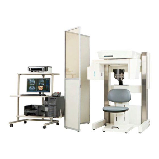
10.0 TECHNICAL SPECIFICATIONS 10.1 General Information MiniCAT™ Scanner System Description MiniCAT™ is a compact, upright, volume computed tomography (VCT) imaging system designed for high-resolution, low-radiation imaging of the sinuses, skull base and temporal bones. System Dimensions 40" L x 36" W x 67" H... - Page 86 FRONT VIEW TOP VIEW © 2014 Xoran Technologies LLC MiniCAT™ User Manual v5.2...
-
Page 87: Imaging Properties
SMOOTH (400x400 120 kVp 7 mA 11.5 ms 10 s (300) 0.4, 0.4, f1) Sinus 10s 12.08 SMOOTH (400x400 120 kVp 7 mA 11.5 ms 10 s (150) 0.4, 0.4, f1) © 2014 Xoran Technologies LLC MiniCAT™ User Manual v5.2... - Page 88 120 kVp 7 mA 11.5 ms 40 s Calibration 0.4, 0.4, f1) Note: If you are using legacy acquisition protocols and are unsure which source you have, please contact Xoran Customer Service. © 2014 Xoran Technologies LLC MiniCAT™ User Manual v5.2...
- Page 89 Sinus 20s (600): 97.0 mGy-cm Sinus 10s (300): 49.2 mGy-cm Sinus 10s (150): 24.6 mGy-cm T Bone 20s (600): 76.9 mGy-cm ISI Source Sinus 40s (600): 85.2 mGy-cm Sinus 20s (300): 42.5 mGy-cm © 2014 Xoran Technologies LLC MiniCAT™ User Manual v5.2...
- Page 90 Temporal Bone: Selectable (0.3 mm minimum reconstructed) Pitch or table feed Scan FOV Sinus: 16 x 10.5 cm Temporal Bone: 16 x 6 cm Gantry tilt/angle Kernel/filter Sinus: Hann Temporal Bone: Ramp © 2014 Xoran Technologies LLC MiniCAT™ User Manual v5.2...
- Page 91 Sinus 20s (600), Sinus 40s (600) 2.88 ± 1.00 Sinus 10s (300), Sinus 20s (300) 4.86 ± 2.18 Sinus 10s (150) 7.09 ± 2.60 T Bone 20s (600), T Bone 40s (600) 5.72 ± 2.19 © 2014 Xoran Technologies LLC MiniCAT™ User Manual v5.2...
- Page 92 (FWHM) resolution of the Slice Sensitivity Profile (SSP). The SSP is obtained by scanning a small, sub-voxel bead in the QC module. However, the nominal tomographic section thickness is fixed for each protocol and is not selectable prior to acquisition. Slice Sensitivity Profiles © 2014 Xoran Technologies LLC MiniCAT™ User Manual v5.2...
- Page 93 Sinus 20s (600), Sinus 40s (600) Sinus 10s (300), Sinus 20s (300) Sinus 10s (150) © 2014 Xoran Technologies LLC MiniCAT™ User Manual v5.2...
- Page 94 The Modulation Transfer Function (MTF) is the amplitude frequency response of the imaging protocol and is a commonly used measure of system blur. The MTF profiles are given in the figures © 2014 Xoran Technologies LLC MiniCAT™ User Manual v5.2...
- Page 95 The phantom was positioned to maximize the view of the axial length of the phantom. More viewable slices translate into more data for estimation and improved MTF estimation accuracy. © 2014 Xoran Technologies LLC MiniCAT™ User Manual v5.2...
- Page 96 Sinus 20s (600), Sinus 40s (600) Sinus 10s (300), Sinus 20s (300) © 2014 Xoran Technologies LLC MiniCAT™ User Manual v5.2...
- Page 97 Sinus 10s (150) T Bone 20s (600), T Bone 40s (600) © 2014 Xoran Technologies LLC MiniCAT™ User Manual v5.2...
-
Page 98: X-Ray Specifications
Pulse Train Time: 40 seconds on, 10 minutes off Less than 5 ms @ 125 kVp 7 mA Rise Time (X-Ray Output) measured @ 50% - 90 % of the voltage waveform. © 2014 Xoran Technologies LLC MiniCAT™ User Manual v5.2... - Page 99 (SourceRay Source) Minimum Permanent Filtration 10.6 mm Al equivalent (ISI Source) Measurement Criteria for Peak 50% - 90 % of the voltage waveform, 125 kVp, 58.8 Voltage, Current, and Pulse Width © 2014 Xoran Technologies LLC MiniCAT™ User Manual v5.2...
-
Page 100: Scatter Map
120 centimeters above the floor (see diagram below). The MiniCAT™ was operated at 120 kVp and 48.3 mAs per Sinus 20s (600) protocol for all secondary radiation measurements reported. © 2014 Xoran Technologies LLC... - Page 101 © 2014 Xoran Technologies LLC MiniCAT™ User Manual v5.2...
- Page 102 The secondary radiation doses were also recorded at 0.5 meters above and 0.5 meters below the horizontal plane intersection the central slice at azimuthal locations A through F and at a radius of 1.0 meter. These measurements are reported in the following tables. © 2014 Xoran Technologies LLC MiniCAT™ User Manual v5.2...
-
Page 103: Dose Information
Sinus 20s (600): 120 kVp, 7 mA, 11.5 ms pulses, 600 pulses (48.3 mAs), 11.7 mm Al equivalent fixed filtration, 0.542 ± 0.048 mm tomographic section thickness. Sinus 10s (300): 120 kVp, 7 mA, 11.5 ms pulses, 300 pulses (24.15 mAs), 11.7 mm Al equivalent © 2014 Xoran Technologies LLC MiniCAT™ User Manual v5.2... - Page 104 Result Subsection (SourceRay Source) (c)(2)(i)(a) CTDI at center for typical factors Sinus 20s (600) 5.4 mGy Sinus 40s (600) CTDI at center for typical factors T Bone 20s (600) 7.7 mGy © 2014 Xoran Technologies LLC MiniCAT™ User Manual v5.2...
- Page 105 Sinus Sinus 40s (600) protocol was found to be at approximately the 8 o’clock position when facing the MiniCAT and looking down on the CTDI phantom. CTDI at max periphery. The location of T Bone 20s (600) 7.1 mGy...
- Page 106 T Bone 40s (600): 125 kVp, 7 mA, 18 ms pulses, 600 pulses (75.6 mAs), 10.6 mm Al equivalent fixed filtration, 0.532 ± 0.043 mm tomographic section thickness. The nominal tomographic section thickness is fixed for each protocol and is not selectable prior to © 2014 Xoran Technologies LLC MiniCAT™ User Manual v5.2...
-
Page 107: Installation Requirements
(90° from max) (180° from max) (270° from max) T Bone 40s (600) 10.6 Installation Requirements Room Requirements Recommended Room Size 8' x 10' Internet Connectivity High speed connection is required for service/maintenance. © 2014 Xoran Technologies LLC MiniCAT™ User Manual v5.2... - Page 108 Note: The operator of the MiniCAT™ must be able to make visual and audio contact with the patient being scanned while behind radiation barrier. Power Japan Japan Europe Requirement ~200V ~100V ~220-240V ~115V Line Voltage 50-60 Hz 50-60 Hz 50-60 Hz...
- Page 109 MiniCAT™ scanner hardware or workstation/server. Installation/Relocation The MiniCAT™ cannot be installed or relocated by the user. Proper and safe setup requires specialized training and must be performed by a Xoran Technologies Customer Service Engineer. Call Xoran 1-800-70-XORAN (800-709-6726) for assistance.
- Page 110 Administrative Station or a Physician's Office, where a laptop computer may be used, it is recommended that a minimum of additional 3rd Party Software be installed and that anti-virus software be utilized. Xoran cannot assure that the Minimum Requirements, outlined below, will allow 3D Viewing to run optimally as intended.
- Page 111 OpenGL and 3D textures. Depending on the video card model and driver implementation, this General Minimum Specification might not be sufficient for 3D to work as intended. Xoran does not assure 3D Viewing will work, as intended to, even when the...
-
Page 112: Igs Compatibility
CD or DVD. Fast network card is required (100Mb/s or more). Network The computer should be able to reach Xoran dedicated router through local network. USB port An available USB port is required for HASP key provided by Xoran. - Page 113 Medtronic Fusion BrainLAB (Requires Pac Xfer Version 5.2 or Higher) Platform Compatible with Xoran VectorVision VectorVision2 VectorVision Compact (for ENT) Kolibri Stryker Platform Compatible with Xoran Stryker Navigation System (original) NavII mNAV © 2014 Xoran Technologies LLC MiniCAT™ User Manual v5.2...
-
Page 114: Quality Control Tests
Xoran provides a quality control phantom to use for these quality control tests. Xoran has developed these procedures to scan only the sinuses and temporal bones, which are high- contrast or ‘bone window’ imaging tasks that require isotropic high spatial resolution and do not require the level of contrast resolution needed for other imaging tasks, such as brain or muscle tissue scanning, etc. - Page 115 Image Noise Annually 11.9 Nominal Tomographic Section Thickness and Annually Slice Sensitivity Profile Note: The required frequency of QC tests may vary according to state regulations, accrediting organizations, or your medical physicist. © 2014 Xoran Technologies LLC MiniCAT™ User Manual v5.2...
- Page 116 Fig. 5. Top Left: the QC Phantom and the Custom QC and Uniformity Material modules in the MiniCAT. Top Right: Photo of the QC Module. Bottom Left: Simplified drawing of the QC Module. Bottom Right: Coronal and Axial images of an MiniCAT scan of the QC phantom.
-
Page 117: Laser Accuracy
Press button once to turn on the laser for a few seconds. Press and hold for three seconds to turn on the laser for five minutes. Side lasers should align with the alignment stickers on perimeter of X-ray detector. © 2014 Xoran Technologies LLC MiniCAT™ User Manual v5.2... -
Page 118: Ct Number Of Reference Materials
2. Draw a circular region entirely within each of the reference material boundaries. Each circular region should range in size from 65-80 square millimeters. 3. Record the mean value for each material and compare with the acceptable range for each. Tolerance: © 2014 Xoran Technologies LLC MiniCAT™ User Manual v5.2... -
Page 119: Spatial Resolution
-200 to 0 HU 11.3 Spatial Resolution Equipment and Scan: Quality Control Phantom, Resolution QC scan High Contrast Procedure: 1. Using the Resolution QC scan, position the Quality Control phantom at the central slice. © 2014 Xoran Technologies LLC MiniCAT™ User Manual v5.2... - Page 120 (see picture below). The picture below is an example of a typical window level for accurate lp/cm viewing. © 2014 Xoran Technologies LLC MiniCAT™ User Manual v5.2...
-
Page 121: Ct Number And Standard Deviation Of Water
Tolerance: Greater or equal to 4 targets should be distinguishable. 11.4 CT Number and Standard Deviation of Water Equipment and Scan: Quality Control Phantom, Water Calibration protocol. © 2014 Xoran Technologies LLC MiniCAT™ User Manual v5.2... -
Page 122: Artifact Test
(Ensure that no rotations of the data have been applied – i.e.: restore original tilts.) The image below represents artifacts that are visible in a reconstruction. This may indicate the need for offset and gain calibration. © 2014 Xoran Technologies LLC MiniCAT™ User Manual v5.2... -
Page 123: Uniformity Test
11.6 Uniformity Test Equipment and Scan: Quality Control Phantom, Water QC scan Procedure: 1. Scroll axially approximately 2 cm above the QC Module to the large section of the Water Module. © 2014 Xoran Technologies LLC MiniCAT™ User Manual v5.2... -
Page 124: Contrast Scale
3. Acquire a Scout for T Bone 20s (600). Label this scan “Contrast Scan 1” as well. 4. Acquire a Scout for Sinus 20s (600), Sinus 10s (300), Sinus 10s (150). Label these scans “Contrast Scan 2.” © 2014 Xoran Technologies LLC MiniCAT™ User Manual v5.2... - Page 125 Central slice [pixel] value. 15. Using the Distance Tool, draw a vertical line centered horizontally across the scout. 16. Create a circular region of 100 mm (± 20 mm ) centered at the crosshairs. © 2014 Xoran Technologies LLC MiniCAT™ User Manual v5.2...
- Page 126 User QC Spreadsheet. If the water attenuation value corresponds to more then one energy (multiple table rows), record the mean value of the Teflon-Water Differential. Attenuation units: [cm Energy Water Teflon Teflon-Water © 2014 Xoran Technologies LLC MiniCAT™ User Manual v5.2...
- Page 127 0.186 0.196 0.38 0.184 0.195 0.376 0.181 0.194 0.374 0.18 0.193 0.372 0.179 0.192 0.369 0.177 0.192 0.367 0.175 0.191 0.365 0.174 0.19 0.363 0.173 0.189 0.361 0.172 0.189 0.359 0.17 © 2014 Xoran Technologies LLC MiniCAT™ User Manual v5.2...
- Page 128 26. Open the Sinus 20s (600) reconstruction, and locate the central slice (click Study Details. The central slice is the opposite of zmin). Draw a 65-80 mm 2 circular region within the Teflon material insert. © 2014 Xoran Technologies LLC MiniCAT™ User Manual v5.2...
- Page 129 Section of the User QC Spreadsheet. 30. Repeat Steps 26-29 for Sinus 10s (300), Sinus 10s (150), and T Bone 20s (600). Tolerance: Sinus 20s (600): 0.00023 to 0.00034 Sinus 10s (300): 0.00021 to 0.00041 © 2014 Xoran Technologies LLC MiniCAT™ User Manual v5.2...
-
Page 130: Image Noise
Spreadsheet. Note that Contrast Scale must be performed before the Image Noise test. 3. Repeat these steps for each of the clinical scans. Tolerance: Sinus 20s (600): 1.88 to 3.87 Sinus 10s (300): 2.69 to 7.04 © 2014 Xoran Technologies LLC MiniCAT™ User Manual v5.2... -
Page 131: Nominal Tomographic Section Thickness And Slice Sensitivity Profile
QC module. Specifically, the custom QC module contains a 0.280 mm tungsten bead in the upper half of the phantom. Note: See below picture as reference. Procedure: © 2014 Xoran Technologies LLC MiniCAT™ User Manual v5.2... - Page 132 Sinus 10s (300): 0.476 to 0.618 Sinus 10s (150): 0.406 to 0.665 T Bone 20s (600): 0.489 to 0.574 Note: Failure to be within these ranges implies poor geometric calibration or mechanical problems with the device. © 2014 Xoran Technologies LLC MiniCAT™ User Manual v5.2...
-
Page 133: Index
CUSTOMER SERVICE (800) 709-6726 international +1 (734) 418-5100 5210 South State Rd. Ann Arbor, MI 48108 info@xorantech.com www.xorantech.com...
Need help?
Do you have a question about the xoran and is the answer not in the manual?
Questions and answers