Sign In
Upload
Manuals
Brands
Olympus Manuals
Microscope
Fluoview-1000
Olympus Fluoview-1000 Manuals
Manuals and User Guides for Olympus Fluoview-1000. We have
1
Olympus Fluoview-1000 manual available for free PDF download: User Manual
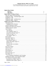
Olympus Fluoview-1000 User Manual (50 pages)
Brand:
Olympus
| Category:
Microscope
| Size: 35.78 MB
Table of Contents
Table of Contents
1
Quick Start Guide
5
Start-Up
5
Shut-Down
5
Quick Guide to Getting an Image
6
Figure 1. Main Fluoview Window
6
Finding the Sample - Transmitted Light or DIC
7
Place the Sample on the Microscope
7
Figure 2. Enlarged View of Fluoview Windows
8
Brightfield Vs. DIC
9
Figure 3. Microscope Controller Window
9
Finding the Sample - Epi-Fluorescence
9
Finding the Sample - Confocal
10
Table 1. Mirror Buttons
10
Setting up for Confocal Microscopy
11
Choosing the Dye
11
Figure 4. Image Acquisition Window - Basic Controls for Finding the Image
11
Changing the Dye Selection
12
Confocal Acquisition Controls
12
Table 2. Objective Lenses Available
12
Settings for the Fluorescence Detectors
13
Additional Detector Controls
13
Laser Controls
14
Line Selection
14
Line Availability, Figure 7.2
14
Figure 5. Channel Settings for the Photomultiplier Tube (PMT)
14
Table 3. Scan Speed and Acquisition Time
14
Laser Power Selector, Figure 7.3
15
Scroll List, Figure 7.4
15
Line Settings, Figure 7.5
15
Figure 7. Selecting Laser Lines and Setting Laser Power
15
Scan Controls
16
Dwell Time, Figure 6.2
16
Figure 6. the Scan Mode and Scan Size Settings of the Acquisition Setting Window
16
Scan Mode, Figure 6.1
16
Box Size, 6.5
17
Image Aspect Ratio, Figure
17
Scan Regions, Figure 6.3
17
Autohv, 6.6
18
Box Size Effect on Resolution
18
Scan Area Controls
18
Field of View, 8.1
18
Scan Area
18
Scan Rotation
18
Scan Zoom, 8.4
19
Reset Scan Values, 8.5
19
Scan Offset, 8.6
19
Figure 8. Scan Controls for Zoom, Pan and Rotation
19
Figure 9. Sequential Imaging Control
20
Figure 9A. Sequential Imaging Control
20
Use of Sequential, Simultaneous and Averaging
21
Simultaneous Capture Versus Sequential Capture
21
Two Methods for Sequential Capture
21
Sequential Frame Capture, Figure 9.1
21
Sequential Line Capture, Figure 9.1
21
Scan Mode and Sequential Capture
22
Using Channel Groups, Figure 9A
22
Adjusting the Channels with Sequential Imaging
22
Adjusting the Channels with Simultaneous Imaging
22
Collecting a Z-Series
23
Determine the Depth of the Optical Volume
23
Which Way Does the Focus Move
23
Figure 10. Z-Series Control Window
24
Figure 11. Z-Series Control Window
24
Settings for Z-Series Collection
25
What Is Axial Resolution
25
Vertical Sections
26
Files: Saving and Transferring
26
Saving Files
26
The Olympus "OIB" Format
26
The Olympus "OIF" Format
26
Where Are Your Files on the Confocal
27
Export File Types
27
Add a Scale Bar to Your File
27
Opening Your Files
27
Olympus Free File Viewer
28
Offline Fluoview Workstation
28
Importing Olympus Datasets into Imagej
28
Loci-Tools Plugin for Imagej
28
Import Channel Order Macro
28
Image 5D Plugin
28
File Import from the Oif.files Directory
29
File Transfer
29
File Transfer to Mac Pro 'Fluoview', Confocal File Server
29
Saving Files Directly to Mac Pro 'Fluoview
29
USB and Firewire Ports on the Mac Pro
29
Recording to CD-ROM and DVD-ROM on the Mac Pro
30
Networked File Transfer to Remote Servers from the Fluoview Mac Pro
30
Using FTP to Windows Computers
30
Remote Access to the Mac Pro from Your Laboratory
30
Optimizing Image Collection
31
Qualities of an Optimal Confocal Image
31
Overview of Image Capture Properties
31
Overview of Basic Image Capture
31
Basic Settings for Confocal Microscopy
31
Labeling Controls
32
Negative Controls
32
Positive Controls
32
The Confocal Aperture
32
Optimal Iris Diameter
32
Bleedthrough
33
Reducing Bleedthrough
33
Procedure to Minimize Bleedthrough
33
Noise
34
Signal Noise (Shot Noise)
34
Detector Noise
35
Kalman Averaging
35
Background
35
Bright Images Versus Scorching the Specimen
35
Images with Weak Signals
35
Using Zoom
36
Zoom Reduces Objective Lens Aberrations
36
Use Zoom to Fulfill Sampling Requirements for the Specimen
36
Use Zoom to Fulfill Adequately Sample the Objective
36
Obtaining a Z-Series
37
Resolution
37
Table 4. Optimal Z-Step Values
37
Box Size
38
Considerations for Increasing Resolution
38
Sampling Frequency and the Nyquist Theorem
38
Table 7. Laser Lines on the FV-1000
38
Zoom
38
Size of Pixels
39
Table. 5 Pixel Size (ΜM) Versus Box Size and Magnification at Zoom = 1
39
Figure 12. DIC and Focus Controls, Lower Half of the Microscope
40
Microscope Controls
41
Focus
41
Fine/Coarse Focus
41
Lowering the Objectives
41
Manual Shutter
41
Differential Interference Contrast (DIC)
41
DIC Analyzer
41
DIC Polarizer
42
DIC Mirror
42
Setting Koehler Illumination
42
Figure 13. Condenser and DIC Controls
42
Condenser Position
43
Figure 14. Tilting the Condenser
43
Photoactivation Methods
44
Zoom Versus Laser Power
44
Recording Initial Recovery Event
44
Methods to Photoactivate/Bleach
44
Using the Stimulus Window
44
Figure 15. the Stimulus Setting Window for Photoactivation
45
Avoid Spontaneous Photoactivation
46
Controlling Computer Display Brightness
46
Figure 16. Photobleaching an ROI
46
Set 'Darkroom' Color Mode
47
Figure 17. the Toolbar Item for Control over Display Color
47
Figure 18. Fluoview Appearance in Darkroom Color
47
Technical Information
48
Caring for Objectives
48
FV-1000 Optical Specifications
48
Table 6. Dichroic Mirrors and Barrier Filters
49
Table 8. Index of Refraction for Common Mounting Materials
49
No Image
50
Start-Up Problems
50
Scans, but no Image
50
The Confocal Image Appears Unevenly Illuminated
50
The Epifluorescent or Tungsten Images Appear Unevenly Illuminated
50
Advertisement
Advertisement
Related Products
Olympus FLUOVIEW FVMPE-RS
Olympus FLUOVIEW FV300
Olympus Fluoview FV1000
Olympus FH
Olympus FHT-521
Olympus FHT-522
Olympus FHT-523
Olympus FHT-533
Olympus FHT-523-SW
Olympus FV3000
Olympus Categories
Digital Camera
Voice Recorder
Microscope
Camera Accessories
Camera Lens
More Olympus Manuals
Login
Sign In
OR
Sign in with Facebook
Sign in with Google
Upload manual
Upload from disk
Upload from URL