Sign In
Upload
Manuals
Brands
AGFA Manuals
Medical Equipment
5530/100
AGFA 5530/100 Manuals
Manuals and User Guides for AGFA 5530/100. We have
1
AGFA 5530/100 manual available for free PDF download: User Manual
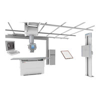
AGFA 5530/100 User Manual (315 pages)
Brand:
AGFA
| Category:
Medical Equipment
| Size: 11.58 MB
Table of Contents
Table of Contents
2
Legal Notice
9
Introduction to this Manual
10
Scope of this Manual
11
About the Safety Notices in this Document
12
Disclaimer
13
Introduction
14
Intended Use
15
Intended User
16
Configuration
17
Applied Parts
19
Options and Accessories
20
Operation Controls
21
Radiographic Table
22
Radiographic Wall Stand
23
Control Panel of the X-Ray Tube Head Unit
24
Remote Control for the X-Ray Tube Head Unit
25
Tube Head Display
27
MUSICA Acquisition Workstation (NX)
28
Software Console
29
Extra Monitor in the Examination Room
30
DR Detector Switch
31
Automatic Positioning Button
32
X-Ray Generator Mini Console (Spellman)
33
X-Ray Generator Mini Console (EMD)
34
Exposure Button
35
Automatic Collimator
36
Collimator Camera
37
Portable DR Detector
39
Emergency Stop Button
40
Emergency Shutdown Power Switch
42
Power off Behaviour
43
Installation
44
HF-Emission and Immunity
44
Radiation Protection
45
Monitoring of Personnel
46
Protected Area and Significant Zones of Occupancy
47
Skin Dose Levels According to IEC 60601-2-54
54
Ongoing Quality Control in Digital Radiography
55
Labels
56
Warning Labels on the Radiographic Table
58
Warning Labels on the Radiographic Wall Stand
58
Type Label
59
DR Detector Identification Label
60
Additional Labeling of the Tube Head Unit
61
Additional Labeling of the Radiographic Table
62
Additional Labeling of the Radiographic Wall Stand
63
Labeling of the Bucky
64
Labeling of the Automatic Exposure Control (AEC)
66
Labeling of the DR Generator Sync Box
67
Labeling of the X-Ray Generator (Spellman)
68
Labeling of the X-Ray Generator (EMD)
70
Labeling of the Remote Control
71
Labeling of Components with Earthquake Resistance
72
About the Software
73
Cleaning and Disinfecting
74
Cleaning
75
Disinfecting
77
Disinfecting Safety Directions
78
Approved Disinfectants
79
Maintenance
80
Maintenance of the Radiographic Table, Radiographic Wall Stand and X-Ray Tube Head Unit
80
Safety Directions
84
General Safety Directions
85
Safety Directions for the X-Ray System
87
Safety Directions for the Radiographic Table
89
Safety Directions for the Ceiling Suspension
90
Basic Workflow
91
Starting the System
92
Automated Workflow for Warming-Up of X-Ray Tube
92
Performing an Exposure Using the DR Detector
94
Step 1: Retrieve the Patient Info
95
Step 2: Select the Exposure
96
Step 3: Prepare the Exposure
97
Step 4: Check the Exposure Settings
98
Step 5: Execute the Exposure
99
Step 6: Perform a Quality Control
100
Performing a Digital Tomosynthesis Examination
101
Step 1: Prepare the Examination
102
Step 2: Position the X-Ray System and the Patient
105
Step 3: Check the Exposure Settings
106
Step 4: Execute the Digital Tomosynthesis Exposure Sequence
107
Step 5: Perform a Quality Control
110
Performing an Exposure Using a CR Cassette
111
Step 1: Retrieve the Patient Info
112
Step 2: Select the Exposure
113
Step 3: Prepare the Exposure
114
Step 4: Check the Exposure Settings
115
Step 5: Execute the Exposure
116
Step 6: Repeat Steps 2 to 5 for the Next Subexposures
117
Step 7: Digitize the Image
118
Step 8: Perform a Quality Control
119
Performing a Full Leg Full Spine Examination
120
Stopping the System
121
Guidelines for Pediatric Applications
122
Software Console and Tube Head Display
124
Action Buttons
126
Planned Exposures
127
Live Camera Image on the Tube Head Display
128
Previewing the Position of the Collimation Area and the AEC Fields
129
Prerequisites for Previewing Collimation and AEC Fields
130
Non-Perpendicular Angles
131
Image Preview Window
132
Main Screen of the Tube Head Display
133
Device Status Frame
134
Ready for Exposure Status
135
Modality Position
136
DR Detector Switch
137
Filter Status
138
Grid Status
139
Radiation Status
140
Unknown Status
141
Generator Controls
142
One Point, Two Point and Three Point Working Modes
143
Radiographic Parameters
145
Focal Spot Indicator
146
Automatic Exposure Control (AEC)
147
X-Ray Tube Load
152
DAP Value
153
Heat Units
154
DR Detector and X-Ray Tube Head Alignment
155
X-Ray Modality Controls
157
Positioning Controls
158
Positioning Screen of the Tube Head Display
159
Actual and Target Position Parameters
160
Collimator Parameters
161
Automatic Positions
162
Tracking the Radiographic Table
167
Tracking the Radiographic Wall Stand
169
Automatic Centering
171
Controls for Digital Tomosynthesis
173
Radiographic Parameters for Digital Tomosynthesis
174
Position Parameters for Digital Tomosynthesis
175
Reconstruction Parameters
176
Adjusting the Reconstruction Settings for Digital Tomosynthesis
177
System Messages
178
Ceiling Suspension
180
Control Panel of the Tube Head Unit
181
Positioning the X-Ray Tube
182
Stop Positions
184
Collision Indicator
185
Positioning the X-Ray Tube Using the Remote Control
186
Automatic Collimator
189
Semi-Automatic Collimation Mode
191
Manual Collimation Mode
192
Collimation Area for Free Exposures
193
Dose Area Product Meter (DAP)
194
Effect of SID on Patient Dose
195
Radiographic Table
196
Positioning the Radiographic Table
197
Positioning the Floating Tabletop
198
Adjusting Height
199
Collision Protection
200
Positioning the Bucky
201
Radiographic Table Accessories
202
Mounting the Patient Hand Grips
203
Mounting the Tabletop Hand Grips
204
Foot Pedals at the Rear Side
205
Mattress
206
Lateral Cassette Holder
207
Compression Belt
208
Radiographic Wall Stand
209
Positioning the Radiographic Wall Stand
211
Radiographic Wall Stand Accessories
213
Patient Hand Grips
214
Mounting the Lateral Arm Rest
215
Spacer
216
Wall Stand Fixation Kit
217
Bucky
218
Bucky Configuration
220
Rotating the Bucky
221
Loading of the Bucky in the Radiographic Table
222
Loading of the Bucky in the Radiographic Wall Stand
223
Unloading of the Bucky in the Radiographic Table
224
Unloading of the Bucky in the Radiographic Wall Stand
225
Opening the Bucky Drawer Configured with an Earthquake Kit
226
Automatic Cassette Size Sensing
227
Bucky Types
228
Cassette and Detector Formats
230
Standard Cassette Formats
231
DR Detector Formats and Orientation
232
Orientation of DR 10S in the Bucky
233
Orientation of DR 14S in the Bucky
234
Orientation of DX-D 10C, DX-D 10G in the Bucky
236
Using DX-D 45C, DX-D 45G, XD 10, XD⁺10 Only Outside the Bucky
237
Grids
238
Anti-Scatter Grids
239
Grid Focal Distance Color Indication
240
Grid Detection
241
Storage Box for DR Detector and Grids
242
Changing the Grid Configured with an Earthquake Kit
243
Automatic Exposure Control (AEC)
244
X-Ray Generator Mini Console
245
Starting and Stopping the Generator
246
X-Ray Tube Start-Up Modes
247
X-Ray Generator Messages and Warning Signals (Spellman)
248
X-Ray Generator Messages and Warning Signals (EMD)
249
Exposure Parameters
250
Radiographic Parameter Limits
252
Exposure Termination
254
Problem Solving
255
Restoring Connection between Generator and NX after Generator Failure
256
Automatic Collimation Always too Wide or too Narrow
257
No Table Movement
259
No Movement When Using Remote Control
260
DR Detector Is Exceeding the Maximum Working Temperature
261
DR Detector Must be Recalibrated
262
Tube Head Display Shows Screen to Check Network Connection
263
Radiographic Parameter Limits
264
Product Information
265
Compatibility
266
Connectivity
267
Compliance
268
General
269
Safety
269
Electromagnetic Compatibility
269
X-Ray Safety
270
X-Ray Accuracy
270
Environmental Compliance
270
Biocompatibility
270
Usability
270
Earthquake Resistance
270
Equipment Classification
271
Patient Data Security
272
Requirements on the Operating Environment
272
Product Complaints
274
Environmental Protection
275
System Documentation
277
Installing the Online Help
277
Training
279
Technical Data
280
DR 600 Technical Data
281
Generator Technical Data (Spellman)
283
Generator Technical Data (EMD)
284
Radiographic Table Technical Data
285
Wall Stand Technical Data
286
Ceiling Suspension Technical Data
288
X-Ray Tube Technical Data
289
Bucky Unit Technical Data
291
Automatic Exposure Control (AEC) Technical Data
293
Automatic Collimator Technical Data
294
Dose Area Product Meter (IBA DAP) Technical Data
295
Dose Area Product Meter (Vacutec DAP) Technical Data
296
Fixed DR Detector
297
Portable DR Detector Technical Data
301
NX Workstation Technical Data
302
DR Generator Sync Box Technical Data
303
Remarks for HF-Emission and Immunity
304
Immunity to RF Wireless Communication Equipment
309
Precautions on EMC
310
Cables, Transducers and Accessories
311
Advertisement
Advertisement
Related Products
AGFA 5520/100
AGFA 5520/200
AGFA 5522/600
AGFA 5523/115
AGFA 5523/210
AGFA 5523/220
AGFA 5523/250
AGFA 5523/265
AGFA 5523/270
AGFA 5523/275
AGFA Categories
Medical Equipment
Printer
Scanner
Digital Camera
Measuring Instruments
More AGFA Manuals
Login
Sign In
OR
Sign in with Facebook
Sign in with Google
Upload manual
Upload from disk
Upload from URL