
Summary of Contents for NDX UNIPICK+
- Page 1 Cell and Tissue Acquisition/Deposition System with Universal Straddle Attachment User Manual Get a Hold of Your Cell 20 20...
-
Page 2: Table Of Contents
Contents Introduction Safety Components and Accessories Initial Set Up Using UnipicK+™ UnipicK+™ Software – Main Controls UnipicK+™ Software – Automation Basic Operations Sample Protocols Troubleshooting Technical Specifications System Performance Warranty and Liability Contact... -
Page 3: Introduction
Introduction UnipicK+ , a capillary-based cell and tissue acquisition system, is the key to reliable, precise, cell and tissue sample acquisition prerequisite for a range of in vitro studies, including cell and region-specific tissue experimentation and single cell analysis. This vacuum-assisted capillary based system is compatible with most inverted microscopes and allows for rapid and efficient acquisition and deposition of specific cells from adherent cell cultures based on their morphology, location or fluorescent label. -
Page 4: Safety
Safety Handle glass capillaries with care. Capillaries have sharp tips and can break on or under skin surface upon contact. It is strongly recommended to always wear goggles and gloves during handling. Install UnipicK+ over an inverted microscope, level surface and allow at least 5 inches of space all around UnipicK+ Always use the power supply and cord provided with the unit. -
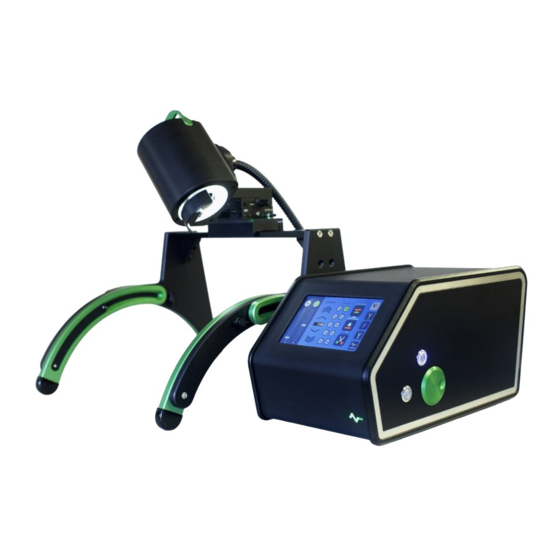
Page 5: Components And Accessories
Components and Accessories UnipicK+ (Figure 1) comes equipped with a Universal Straddle that may be fitted over most inverted microscopes, UnipicK+ unit (Sampler Head at top with adjustable LED ring light, Vacuum Line and Electric Cables, and Control Box with vacuum and pressure module), and a power supply with cord. -
Page 6: Initial Set Up
Initial Set Up All necessary components are included in the packing. Make sure that the following items have been received upon opening the package. UnipicK+ unit (Head and Control Box) (1) System Alignment Tool (1) Universal Straddle Stand with a bullseye level, a pair of gussets, and short and long horizontal bars (1) Hex wrenches (2, 2.5, 3, 4, &... - Page 7 Assemble the Universal Straddle by attaching a bridge (short or long) to the curved legs with a pair of gussets and screws provided with the unit. DO NOT TIGHTEN THE SCREWS AT THIS TIME. There are possible vertical positions where the bridge can be attached. Choose the appropriate height depending on your microscope.
- Page 8 screws with wingnuts and slide each of the legs down until they all rest on the benchtop. Once the legs are down on both sides, tighten the screws to secure the legs in place. Looking down onto the System Alignment Tool, visually align the center opening on the Tool to the microscope objective...
-
Page 9: Using Unipick
Using UnipicK+™ 5.1 Terminology Starting position: Highest vertical position for the DCU; the initial position when the unit is first turned on and capillary has been mounted or after it is reset. Home position: Calibration of Collecting/Dissecting or Dispensing position of the DCU tip determined by the user. - Page 10 5.3 Filter and DCU attachment Turn the UnipicK+ head 90˚ counterclockwise to expose the male luer connector (Figure 3). Holding the outer rim of the filter, connect the female luer lock connector of the filter to the male luer connector on the UnipicK+ Head.
- Page 11 5.7 Vacuum level and duration control The vacuum or pressure levels may be controlled from 0 to maximum of 70 kPa and the impulse duration may be set from 100 ms to maximum of 1 s (see UnipicK+ Software section). Depending on the sample and the desired result, various vacuum/pressure levels and impulse duration may be used.
- Page 12 Figure 3: UnipicK+ sample collection Head (1). The following components are shown: (2) Shield covering green horizontal calibration light used for DCU tip identification during calibration procedure; (3) Vacuum line and electrical cable. The Head should be turned full 90˚ for disposable capillary (DCU) attachment/removal.
-
Page 13: Unipick+™ Software - Main Controls
UnipicK+™ Software - Main Controls UnpicK+™ software operates the instrument via the touch screen. It offers controls for calibration sequences, sample collection, dispensing, fixed tissue microdissection and single cell adhesion force measurements. The following section describes the main menus, operations, icons and their function. - Page 14 The following controls/icons are present on the right side of the Calibration window: Acquisition plane icon sets the plane where sample collection is to take place; Dispensing plane icon sets the plane where collected sample(s) will be deposited; The Retract feature remembers the most recent Home (Acquisition or Dispensing plane) position.
- Page 15 Auto-dispensing icon takes you to the plate set up and auto-dispensing menu (Available for instruments equipped with a motorized stage. Please contact NeuroInDx representative for details.) When activated (turned green) this icon indicates that auto-dispensing process is ON (Available for instruments equipped with a motorized stage. Please contact NeuroInDx representative for details.) NOTE: Acquisition plane may be changed by bringing the DCU to the desired location using the Green calibration wheel (Figure 2-5) then pressing the wheel.
- Page 16 6.5 Fixed Tissues This window provides manual automatic pulse modes for acquisition of ROIs from fixed tissues such as formalin fixed paraffin embedded or PFA fixed tissue specimen. Pulse is an application of vacuum impulse with predefined duration and strength without lifting of the DCU tip from the tissue surface, which allows mixing the digestion liquid inside the capillary barrel.
- Page 17 6.7 Settings Left half of the screen allows for the selection (from left to right) of main light, calibration light, NDX logo light, and screen light intensities. Three icons on the top right permit selection of DCU vertical movement speed for the fast mode of the calibration wheel.
-
Page 18: Unipick+™ Software - Automation
UnipicK+™ Software – Automation (optional) Auto-collection and auto-dispense features allow the user to automatically collect and dispense multiple samples into PCR tubes. These features require a motorized stage (please contact NeuroInDx representatives for specifications). The process entails Plate setup (7.1), Dispensing parameters selection (7.2), and Target selection (7.3). - Page 19 deselected, and only dark blue wells are remaining for depositing samples. NOTE: Similarly, individual wells may be deselected (tapped once) or prioritized (tapped twice) for the deposition process. NOTE: Before using the auto dispensing function, make sure to set the pressure on the Dispensing window at a minimum of 2kPa.
- Page 20 The progress of deposition will be shown in the auto-dispensing window that may be accessed any time by tapping the green AUTODISP icon. Wells where collected samples were deposited will be indicated with green. For example, the image on the right shows that already four samples were deposited in the first four wells.
- Page 21 The following controls/icons are present on the right side of the Auto-collection window: Preselect icon is used for selection of up to eight targets for auto-collection and deposition Pool icon selects “pool” mode of collection where up to eight preselected targets are collected and dispensed into a single tube Single icon selects “single”...
-
Page 22: Basic Operations
Basic Operations 8.1 Initial Training Exercises with UnipicK+™ For instructional videos visit our website: www.neuroindx.com or YouTube channel: http://www.youtube.com/NDXInc Start UnipicK+ by pressing the Switch on the back panel of the Control box once (green logo and a touch screen will light up). Caution: Wear gloves and protective eyewear when handling DCUs containing glass capillaries. - Page 23 Locate the marking on slide. Using coarse and fine focus adjustment knobs to focus on marking. Carefully bring the DCU down until the annulus of the capillary tip comes into focus as a bright green ring. 10. Stop lowering the DCU when the capillary tip moves with the glass slide when using the mechanical stage.
- Page 24 before lowering the DCU for calibration. The intensity may be adjusted using a sliding bar in the Calibration window menu] Caution: DCUs have sharp tips! Handle with care. Improper handling may cause injuries. Never contact the capillary tip to avoid injury and contamination. Caution: Always use a filter with the DCU to prevent biological material and liquids from entering UnipicK+’s mechanical parts.
- Page 25 tip has made contact. When the DCU tip is in contact with the slide surface, a slight motion of the mechanical stage will result in the simultaneous movement of the capillary tip. If the tug of the slide movement is observed, move DCU tip slightly up (~ few micrometers;...
- Page 26 8.3 DCU Calibration for Adherent Cell Cultures NOTE: Each new DCU must be calibrated before sampling. For instructional video visit our website: www.neuroindx.com or YouTube channel: http://www.youtube.com/user/NDXInc Caution: Wear gloves and protective eyewear when handling DCUs containing glass capillaries. TIP: When working with attached cells it is recommended to wash cells with fresh medium to remove floating cells.
- Page 27 Use the linear stages/micromanipulators (Figure 3) to locate the DCU halo. Use coarse and fine focusing knobs to focus on the cells. Slowly bring the DCU down until the annulus of the capillary tip comes into focus as a green ring. Position the green ring over a clear spot.
- Page 28 Caution: When the UnipicK+ Head is rotated 90˚ counterclockwise, the DCU will not return to its initial Starting position. To return the DCU to the initial position, press the blue Retract button. To avoid breaking DCU tip we recommend returning DCU to its initial Starting position before removing it from the unit.
- Page 29 NOTE: DCU contents may also be released using a syringe with a male luer lock. Make sure that a syringe with a male Luer lock has been prepared with the plunger slightly pulled back and a filter attached to the tip (Figure 5). To empty the contents from the DCU, rotate the UnipicK+ Head 90˚...
- Page 30 Repeat the procedure until the DCU is emptied. 8.6 Using Retract Function to Move Between Wells Press the Retract icon in the Acquisition window. This will bring the DCU up to its original Starting position. Move the plate using the mechanical stage to position the next well under the field of view.
- Page 31 Press the Retract icon in the Calibration menu; this will bring the DCU down to the Standby position. Do not press Acquisition plane icon. The actual DCU tip position may have been changed due to the detachment/attachment procedure. It is safer to recalibrate the DCU.
- Page 32 Figure 5: DCU and filter affixed to a Luer lock sterile syringe. 1 – DCU; 2 – filter, 3 – syringe.
-
Page 33: Sample Protocols
Sample Protocols Protocol 1: Collection of Individual Adherent Cells from Culture Dishes Various adherent cell cultures including human neuroblastoma cell line SH-SY5Y, Chinese hamster ovary (CHO) cells, human melanoma MDA-MB-435 cells and various primary cultures, such as neural progenitors, skin fibroblasts, etc., can be used for the collection of individual cells using both UnipicK and UnipicK+ . - Page 34 When collecting fluorescently labeled cells (Figure 6), calibrate the DCU under bright field as described above, then turn off LED ring light. Turn on the fluorescent illumination to locate desired labeled cells. Turn the LED ring light on and position the cell under the center of the crosshair.
- Page 35 Protocol 2: Sucrose Treated Frozen Brain Tissue Microdissection This protocol describes the isolation of single cells, cell clusters, and subanatomical regions from sucrose treated brain slices (Figure 7). Sucrose treated brain tissue keeps brain morphology intact, and thus ideal for microdissection. Furthermore, this optimized protocol yields high quality RNA from the microdissected material.
- Page 36 NOTE: Optimal vacuum levels and duration can vary depending on the type of samples being dissected. It is recommended that a test slide be used to determine the optimal values of vacuum level and duration prior to sampling. Start at the lowest settings for vacuum level and duration and slowly increase them in turn.
- Page 37 Protocol 3: Microdissection of Native Brain Tissues Both UnipicK and UnipicK+ can dissect live tissues for primary culture and other various downstream molecular analyses. The following is a sample protocol for the collection of live cells from brain tissue to be used for culturing. UnipicK+ can be applied to dissect live cells from other tissues, such as tumors, for primary culture using suitable protocols.
- Page 38 Excise the brain, rinse with cold ACSF, and remove excess liquid with sterile napkin. Keep brain in buffer consisting of Krebs-Ringer solution saturated with carbogen (95% , 5% CO ) on ice until mounted onto glass slides. Glue the brain on the vibratome platform with fast glue (e.g. medical device adhesive Loctide 4014).
- Page 39 17. Plate the cells and incubate at 37°C in a 5% CO atmosphere. Media NB+++ (50 ml) Neurobasal medium 48 ml Penicillin/Streptomycin (100Χ) 500 µl L-glutamine (200 mM), [2 mM final] 500 µl Heparin (5 mg/ml), [2 μg/mL final] 20 µl B-27 (50Χ) 1 ml FGF2 (25 ng/µl), [20 ng/ml final]...
- Page 40 Protocol 4: Microdissection of Microcapillary Cell Walls from Mouse Heart Muscle Tissue This protocol is suitable for dissecting most difficult–to-dissect tissue and isolating high quality RNA from collected samples. Materials: • Liberase DL Research Grade (Roche, REF 05-401-160-001), 5mg vial •...
- Page 41 2. Inhibitor Solution a. 1 proteinase inhibitor tablet in 250ml MEM or PBS Tissue Preparation: Anesthetize the animal with 50 mg/kg Nembutal or another anesthetic approved by your ARC. Flush/perfuse the animal with standard PBS. DO NOT USE ANY FIXATIVES. Remove the heart and other organs and sink them in 15-20% Sucrose in PBS at 4°C overnight.
- Page 42 Prior to removing a DCU from UnipicK+ Head, pull the syringe plunger back and attach a filter to the syringe tip. Carefully remove the DCU from UnipicK+ Head and affix to the filter on syringe. Carefully position the DCU tip into a microcentrifuge tube preloaded with buffer and slowly eject cell/tissue.
-
Page 43: Troubleshooting
10 Troubleshooting If problems with UnipicK+ occur by factors other than manufacturing defects, please review the following guide. If you encounter any other problems, please contact technical support. Problem Cause Solution Increase vacuum pressure and/or duration. Vacuum level is not high enough. -
Page 44: Technical Specifications
11 Technical Specifications Specification Description 1. Illumination Light source 122 LEDs ring light 17.8 in/452 mm (L) x 17.2 in/437 mm (W) plus 9.3 in/237 mm (Control 2. Min Dimensions Box, W) x 24.7in/626 mm (H) 18.8 in/478 mm (L) x 21.1 in/537 mm (W) plus 9.3 in/237 mm (Control 3. -
Page 45: System Performance
12 System Performance Description Specifications Resolution Single Cell Vacuum duration (Ts), seconds 0.1 s to 1.0 s Vacuum strength, kPa Up to 70 kPa Pressure strength, kPa Up to 36 kPa Available DCU IDs, µm From 10 to100 µm Acquisition/Dispensing speed (kPa/Ts), seconds maximum settings (0.1 sec) 1.3 s Maximum settings (1.0 sec) -
Page 46: Warranty And Liability
NDX has made every effort to ensure that this manual is as complete and as accurate as possible, but no warranty or fitness is implied. The information provided is on an “as is” basis. NDX shall have neither liability nor responsibility to any person or entity with respect to any loss or damages arising from the information contained in this manual. - Page 47 NeuroInDx, Inc. SAFETY INFORMATION 20725 S WESTERN AVE, STE 100 Please read the instruction manual carefully in Torrance, CA 90501-1885 order to safely operate any NeuroInDx product Copyright (c) 2016 NeuroInDx, Inc. All rights reserved. NeuroInDx, the NeuroInDx logo, KuiqpicK and UnipicK are trademarks of NeuroInDx, Inc. in the U.S. and/or other countries. Trademarks belonging to the third parties are the properties of their respective owners.