
Subscribe to Our Youtube Channel
Related Manuals for Cortex DermaScan C USB


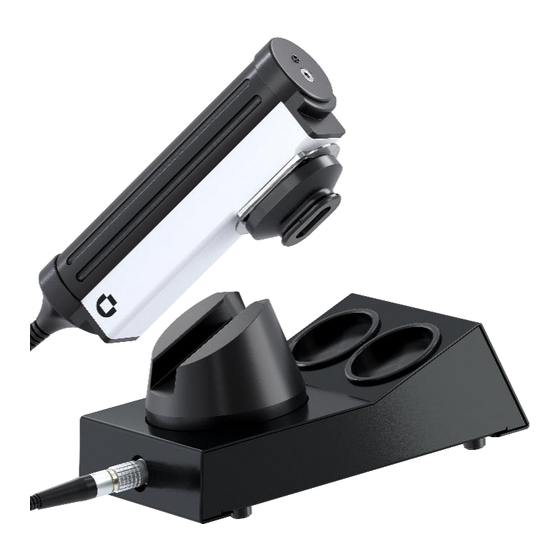
Summary of Contents for Cortex DermaScan C USB
- Page 1 Released 20230914 Instruction Manual Z0300123 UK ® DermaScan C USB Instruction Manual Niels Jernes Vej 6B 9220 Aalborg, Denmark www.cortex.dk cortex@cortex.dk...
- Page 2 ® DermaScan C USB CORTEX TECHNOLOGY...
- Page 3 Copyright © 2023 by Cortex Technology ApS. All rights reserved. No part of this publication may be reproduced, transmitted, stored in a retrieval system, or translated into any language in any form by any means without the permission of Cortex Technology.
-
Page 4: Table Of Contents
® DermaScan C USB CORTEX TECHNOLOGY Contents 1. Symbols ..............................5 2. Warnings ............................... 6 3. Intended use ............................7 4. System overview ........................... 7 4.1. Assembly ............................8 5. 2D scanning probe ..........................9 5.1. General ............................9 5.2. Preparing the probe ........................9 5.2.1. - Page 5 ® DermaScan C USB CORTEX TECHNOLOGY 6.4.1. A-scans........................... 19 6.4.1.1. A-scan measurement toolbar..................... 19 VERTICAL ........................... 20 ARBITRARY ..........................20 CLEAR A-MEAS.......................... 20 END A-MEAS..........................20 6.4.1.2. B-scan measurement toolbar.................... 20 INTENSITY SEGMENTATION ....................22 CLEAR B-MEAS.
-
Page 6: Symbols
® DermaScan C USB CORTEX TECHNOLOGY 1. Symbols Attention, consult accompanying documents. Where applicable, this symbol can be found on the following pages. Type B equipment acc. to EN 60601-1. Class 2 equipment acc. to EN 60601-1. Alternating current, single phase. -
Page 7: Warnings
® DermaScan C USB CORTEX TECHNOLOGY 2. Warnings • Read this entire manual before using or showing to others how to use the DermaScan C USB. ® • Do not use the DermaScan C USB if the device shows visible signs of damage or there is ®... -
Page 8: Intended Use
® USB equipment. Only laptop PC´s delivered by Cortex Technology shall be used with the system. If an additional power supply for the laptop PC is supplied as part of the delivery, this power supply shall newer be used for scanning purposes. It shall only be used, if the PC is disconnected from the DermaScan unit. -
Page 9: Assembly
® DermaScan C USB CORTEX TECHNOLOGY 4.1. Assembly All connectors are marked and/or coded eliminating the risk of incorrect connection of compo- nents. - connect the power supply to the main unit rear connector. - connect the USB cable to a USB port on the laptop PC then to the rear main unit USB connector. -
Page 10: D Scanning Probe
® DermaScan C USB CORTEX TECHNOLOGY 5. 2D scanning probe This chapter applies to all 2D scanning ultrasound probes for the DermaScan C USB irrespective ® of the probe model, scanning frequency and acoustical focusing. 5.1. General The DermaScan C 2D-scanning heads or ‘‘B-probes’’ are scanning in a single plane, the so-called B- mode. -
Page 11: Water Injection
® DermaScan C USB CORTEX TECHNOLOGY Excessive film material and the paper frame is removed by a quick downward pull, which will make the film break over the edge of the transparent acrylic front piece, fig. 2b. Fig. 2b. Mounting the water barrier. -
Page 12: Probe Maintenance
® DermaScan C USB CORTEX TECHNOLOGY Arrange a container to collect the water coming from the scanning head. After removing the black oval ring from the scanning head, the film should be removed by gently pulling the acrylic nose out of the black oval ring. -
Page 13: Operation
® DermaScan C USB CORTEX TECHNOLOGY 6. Operation 6.1. Short form overview 6.1.1. Elements of the main screen Toolbar Ultrasound image Property field Fig. 6. Main screen. Toolbar The toolbar is divided in three sections for controlling the scanning, A-scan measurements and B- scan measurements respectively. -
Page 14: Zoom And Scroll
® DermaScan C USB CORTEX TECHNOLOGY Property field The property field provides information about actual scan settings, settings for already recorded images as well as measured parameters. When making adjustments to settings or when performing measurements, the property field is automatically updated to reflect these actions. -
Page 15: How To Make A Good Image
® DermaScan C USB CORTEX TECHNOLOGY Fig. 8 and 9 shows examples of less reflective material. Vein Nevus Fig. 8. Normal skin over vein. Fig. 9. Intradermal nevus. 6.2.3. How to make a good image Gain setting. Most importantly, the amplification of the signal should be set correctly. As the focused sound beam travels through the tissue it gets attenuated. - Page 16 ® DermaScan C USB CORTEX TECHNOLOGY from the probe front and the skin surface. Re-apply fresh gel and move the probe to a less hairy scanning area. If necessary, hairs may be shaved off the surface (right image). Fig. 14. Gel layer thickness.
-
Page 17: Considerations
® DermaScan C USB CORTEX TECHNOLOGY Fig. 17. High attenuation. The properties of the skin var- ies all over the body and be- tween individuals. In particu- lar areas like the palm, the un- derside of the fingers and sole develop very thick and highly attenuating skin. -
Page 18: Start/Stop Scanning
® DermaScan C USB CORTEX TECHNOLOGY It is of utmost importance to keep a fixed and well-defined thickness of the gel layer between the skin surface and the plastic membrane of the probe. Accordingly, the procedure for applying the gel (section 5.2.3) should be followed closely. -
Page 19: Session
® DermaScan C USB CORTEX TECHNOLOGY SESSION New Session: click to clear all image positions simultaneously. Unsaved im- ages will be permanently erased. Clear Current Position: click to clear the currently active image position. An unsaved image will be permanently erased. -
Page 20: Image Analysis
® DermaScan C USB CORTEX TECHNOLOGY 6.4. Image analysis 6.4.1. A-scans. An ultrasound image is composed by a number of so-called A-scan lines to form a cross-sectional image – the B-scan image. It is beyond the scope of this manual to provide a detailed explanation of the terms A-scan and B- scan, but the reader is encouraged to seek additional literature, which deals with these terms in depth. -
Page 21: Vertical
® DermaScan C USB CORTEX TECHNOLOGY HORIZONTAL Click to perform horizontal (in-depth) measurements. Move the cursor over the image, and a hori- zontal straight line will indicate the actual position within the cross-sectional from which the A- scan curve originates. The A-scan line number (0 – 223) is indicated at the bottom of the screen. - Page 22 ® DermaScan C USB CORTEX TECHNOLOGY EDGE DETECTION Edge detection is used to calculate the distance between two automatically detected lines – e.g. when measuring the average skin thickness over the full scan length. Fig. 25. Edge detection toolbar. Edge detection is associated with a setting of the detection sensitivity relative to the captured A- scans –...
-
Page 23: Intensity Segmentation
® DermaScan C USB CORTEX TECHNOLOGY To facilitate edge detection of the full vertical length the pointer will snap to the bottom/top edge of the image, however, the edge detection may be started from any vertical position of the pointer. -
Page 24: Clear B-Meas
® DermaScan C USB CORTEX TECHNOLOGY Fig. 28. ROI toolbar. REGION OF INTEREST (ROI) Instead of performing a full segmentation within pre-defined borderlines as above, it is possible to select individual areas, so-called regions of interest (ROI), for which the size and mean intensity is calculated. -
Page 25: Maintenance
® DermaScan C USB CORTEX TECHNOLOGY 7. Maintenance No preventive maintenance is needed. The main unit contains no user serviceable parts inside and shall not be opened by unauthorized personnel. 8. Trouble shooting Error Possible cause Solution No external power on laptop PC... -
Page 26: Declaration Of Conformity
® DermaScan C USB CORTEX TECHNOLOGY 9. Declaration of Conformity EC – DECLARATION OF CONFORMITY We hereby declare that the products listed below conform to the requirements of “DI- RECTIVE 2004/108/EC OF THE EUROPEAN PARLIAMENT AND OF THE COUNCIL of 15 December 2004 on the approximation of the laws of the Member States relating to elec- tromagnetic compatibility and repealing Directive 89/336/EEC”...
Need help?
Do you have a question about the DermaScan C USB and is the answer not in the manual?
Questions and answers