Table of Contents
Advertisement
Quick Links
Advertisement
Table of Contents

Subscribe to Our Youtube Channel
Related Manuals for KERN Optics OCM 161


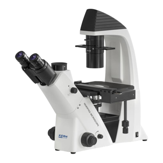
Summary of Contents for KERN Optics OCM 161
- Page 1 KERN & Sohn GmbH Ziegelei 1 Tel: +49-[0]7433- 9933-0 D-72336 Balingen Fax: +49-[0]7433-9933-149 E-mail: info@kern-sohn.com Internet: www.kern-sohn.com User instructions Biological inverted microscope KERN OCM-1 OCM 161, OCM 165, OCM 166, OCM 167, OCM 168 Version 1.1 10/2022 OCM-1-BA-e-2211...
-
Page 2: Table Of Contents
KERN OCM-1 Version 1.1 10/2022 User instructions Biological inverted microscope Table of contents Before use ..................3 General notes ..........................3 Notes on the electrical system ....................3 Storage ............................4 Maintenance and cleaning ....................... 5 Nomenclature ................6 Technical data / Features ............8 Assembly .................. -
Page 3: Before Use
1 Before use 1.1 General notes You must open the packaging carefully, to make sure that none of the accessories in the packaging fall on the floor and get broken. In general, microscopes should always be handled carefully because they are sensitive precision instruments. -
Page 4: Storage
Under no circumstances should you touch the halogen bulb (for transmitted illumination) or the HBO lamp (OCM 165, OCM 166) for the reflected light unit either during operation or directly after use. These bulbs produce significant heat and therefore there is a risk that the user could be severely burnt. So before handling the bulbs, you must check that they have cooled down. -
Page 5: Maintenance And Cleaning
1.4 Maintenance and cleaning In any event, the device must be kept clean and dusted regularly. If any moisture should be occur, before you wipe down the device you must ensure that the mains power is switched off. When glass components become dirty, the best way to clean them is to wipe them gently with a lint-free cloth. -
Page 6: Nomenclature
2 Nomenclature OCM-1-BA-e-2211... - Page 7 OCM-1-BA-e-2211...
-
Page 8: Technical Data / Features
3 Technical data / Features OCM 161 Product dimension: 304×599×530 mm Packing dimensions: 660x590x325 mm Net weight: 13,5 kg Gross weight: 18 kg OCM 165 / 166 / 167 / 168 Product dimension: 304×782×530 mm Packing dimensions: 1050x590x330 mm Net weight:... - Page 9 OCM-1-BA-e-2211...
-
Page 10: Assembly
4 Assembly OCM-1-BA-e-2211... -
Page 11: Objectives
4.1 Objectives The nosepiece must be in its lowest position so that the objectives [1] can be screwed into it. You can then pass the objectives through the opening of the specimen stage and screw them into the nosepiece, so that when you turn the nosepiece in a clockwise direction, the objective with the next strongest magnification appears. -
Page 12: Specimen Stage
4.3 Specimen stage The supplied stage plate [1] needs to be fitted to the opening of the specimen stage, in order to get a support area for relatively small observation objects and to protect the objectives which are located underneath. Furthermore you can attach an object holder to one of the threads on stage surface [2]. -
Page 13: Condenser
4.4 Condenser The condenser [2] must be mounted on the microscope housing underneath the lamp housing. By using the Allen screw [1] it must be firmly attached onto the connection point. The condenser includes the following control elements: Color filter slide (see 5.7 Adjusting the illumination) Aperture diaphragm (see 5.7 Adjusting the illumination) Slot for phase contrast slide (see 8.2 Phase contrast unit) For the connection of a microscope camera and the using of phase contrast or... -
Page 14: Operation
5 Operation 5.1 Getting started The very first step is to establish a power connection using the mains plug. You should first adjust the dimmer to a low level, so that when you look through the eyepiece for the first time, your eyes are not immediately subject to a high level of light. You can now switch on the lighting using the main switch. -
Page 15: Pre-) Focussing
5.2 (Pre-) focussing When you are observing an object, you must have the correct distance to the objective to achieve a sharp image. In order to find this distance at the beginning (without other default settings of the microscope) place the objective with the lowest magnification in the beam path, look through the right eyepiece with the right eye and turn it slowly using the coarse adjustment knob. -
Page 16: Adjusting The Interpupillary Distance
5.3 Adjusting the interpupillary distance With binocular viewing, the interpupillary distance must be adjusted accurately for each user, in order to achieve a clear image of the object. While you are looking through the eyepieces, use your hands to hold the righthand and lefthand tube housing firmly. -
Page 17: Adjusting The Magnification
5.5 Adjusting the magnification After prefocussing has been carried out using the objective with the lowest magnification (see section 5.2) you can then adjust the overall magnification using the nosepiece, as necessary. By turning the nosepiece you can bring any one of the four other objectives into the beam path. -
Page 18: Using Eye Cups
5.6 Using eye cups The eye cups supplied with the microscope can basically be used at all times, as they screen out intrusive light, which is reflected from light sources from the environment onto the eyepiece, and the result is better image quality. But primarily, if eyepieces with a high eye point (particularly suitable for those who wear glasses) are used, then it may also be useful for users who don’t wear glasses, to fit the eye cups to the eyepieces. -
Page 19: Adjusting The Illumination
5.7 Adjusting the illumination To make sure that perfect image results are achieved during microscopic observation, it is important that the direction of light of the microscope is optimised. The following components of the transmitted illumination can be adjusted according to the application requirements. -
Page 20: Changing The Bulb
6 Changing the bulb You must not attempt to change the bulb immediately after the microscope has been used, as the bulb will still be hot and so there is a risk that the user could be burnt. Before changing the bulb the device must be switched off and unplugged. To change the bulb the cover of the lamp housing [b] needs to be removed. -
Page 21: Changing The Fuse
7 Changing the fuse The fuse housing is on the rear of the microscope below the mains power supply socket. With the device switched off and unplugged, you can pull out the housing. When doing this, it is helpful to use a screwdriver or similar tool. The defective fuse can be removed from its housing and be replaced with a new one. -
Page 22: Using Optional Accessories
8 Using optional accessories 8.1 Camera connection Due to the trinocular tube it is possible to connect microscope cameras to the device, in order to digitally record images or sequences of images of an object being observed. After the plastic cover has been removed from the camera adapter connector on the top of the microscope head, then a suitable adapter must be fitted. -
Page 23: Phase-Contrast Units
8.2 Phase-contrast units The standardly supplied phase contrast unit of the microscopes of the series OCM-1 consists of a PH objective (20x), a PH slide, a centring telescope and a green filter. Optionally PH objectives with a magnification of 10x or 40x are available. To use this, you need to place the PH objective into the beam path and push the PH slide (the writing facing up) into the appropriate slot on the condenser. - Page 24 In the field of view you will now see the image of a white (PH slide) and a black (PH objective) ring. The black one is central and the white one possibly is pushed to one side (see left illustration). g.
-
Page 25: Fluorescence Reflected Light Unit
8.3 Fluorescence reflected light unit OCM 165 / 166 / 167 / 168 There are samples, which can be excited by light beams and thereby show emissions, which have different wave lengths than the previous excitation beams. The wave length of the emission is always bigger than the wave length of the excitation (Stokes shift). - Page 26 Overview of wave lengths for excitation and emission per excitation filter Blue line: Wave length excitation Green line: Wave length emission OCM-1-BA-e-2211...
- Page 27 OCM-1-BA-e-2211...
- Page 28 OCM-1-BA-e-2211...
- Page 29 Nomenclature OCM-1-BA-e-2211...
- Page 30 Nomenclature (FL filter unit B / G: OCM 165, OCM 167) Fluorescence filter slide Beam path lock Nomenclature (FL filter unit B /G / V / UV: OCM 166, OCM 168) Beam path lock Fluorescence filter slide OCM-1-BA-e-2211...
- Page 31 Nomenclature (FL unit middle part: OCM 165 / 166 / 167 / 168) Field diaphragm Connection point for lamp housing Nomenclature (FL HBO lamp housing: OCM 165 / 166) OCM-1-BA-e-2211...
- Page 32 Nomenclature (Power supply unit for HBO lamp: OCM 165 / 166) Nomenclature (FL LED lamp housing: OCM 167) Condenser adjustment LED lamp slide OCM-1-BA-e-2211...
- Page 33 Nomenclature (FL LED lamp housing: OCM 168) LED lamp Condenser adjustment adjustment wheel Nomenclature (Power supply unit for LED lamp: OCM 167 / 168) Dimmer Main switch OCM-1-BA-e-2211...
-
Page 34: Assembly And Operation Hbo Versions: Ocm 165 And Ocm 166
8.3.1 Assembly and operation HBO versions: OCM 165 and OCM 166 Assembly In order to put the fluorescence reflected light unit into operation, the following steps have to be carried out. 1. The main body of the FL unit is already firmly connected with the microscope housing and towers above the rear side of the microscope. - Page 35 Control elements for illumination The following control elements for the illumination play a role within the fluorescence microscopy: • Field diaphragm, condenser: Optimisation of the contrast and the light yield Important warnings for the usage of a HBO lamp • During operation the lamp is showing a strong heat development. It should be avoided to come into contact with the lamp housing while and after some time of running the reflected light unit.
- Page 36 Centring the lamp (HBO) When running the HBO lamp for a long time there is the possibility, that the holder of the lamp is moving out of its centre due to the strong heat development. This circumstance has to be corrected when it appears, because therefore the field of view is no longer equally illuminated.
- Page 37 The new installed spare lamp may under no circumstances be touched with naked hands. Impurities will benefit the risk of explosion during operation. If there should be nevertheless impurities, the lamp has to be cleaned. We recommend for this purpose to use a lint-free cloth soaked with an ether alcohol mixture (Proportion: 70/30).
-
Page 38: Assembly And Operation Led Versions: Ocm 167 And Ocm 168
8.3.2 Assembly and operation LED versions: OCM 167 and OCM 168 Assembly In order to put the fluorescence reflected light unit into operation, the following steps have to be carried out. 1. The main body of the FL unit is already firmly connected with the microscope housing and towers above the rear side of the microscope. - Page 39 Control elements for illumination The following control elements for the illumination play a role within the fluorescence microscopy: • Field diaphragm, condenser: Optimisation of the contrast and the light yield Changing the fuse (if existing) The fuse housing is on the rear of the power supply unit below the mains power supply socket.
-
Page 40: Trouble Shooting
9 Trouble shooting Problem Possible causes The mains plug is not correctly plugged in There is no power at the socket The bulb does not light Defective bulb Defective fuse The bulb blows immediately The specified bulb or fuse has not been used The aperture diaphragm and/or field diaphragm are not opened wide enough The selector switch for the beam path is set... - Page 41 Problem Possible causes The aperture diaphragm is not opened wide enough The objective does not belong to this Blurred details microscope Bad image The front lens of the objective is dirty Bad contrast The condenser is not correctly centred Vignetted field of view Dirt / dust on the objective Dirt /dust on the front lens of the condenser Dirt / dust on the eyepieces...
-
Page 42: Service
10 Service If, after studying the user manual, you still have questions about commissioning or using the microscope, or if unforeseen problems should arise, please get in touch with your dealer. The device may only be opened by trained service engineers who have been authorised by KERN.
Need help?
Do you have a question about the OCM 161 and is the answer not in the manual?
Questions and answers